 Indietro
DNA Replication: Mechanisms and Key Enzymes
Guida di studio - Note intelligenti
 Appunti personalizzati basati sui tuoi materiali, ampliati con definizioni chiave, esempi e contesto.
DNA Replication: A Closer Look
Overview of DNA Replication
DNA replication is a fundamental biological process that ensures the accurate copying of genetic material before cell division. It is carried out by a coordinated team of enzymes and proteins, and occurs in both prokaryotic and eukaryotic cells with remarkable speed and fidelity.
Speed and Accuracy: E. coli can replicate its 4.6 million nucleotide pairs in less than an hour, while human cells replicate 6 billion pairs in a few hours, with an error rate of only one per 10 billion nucleotides.
Enzyme Team: More than a dozen enzymes and proteins participate in the process.
Similarity: The basic mechanism is conserved between prokaryotes and eukaryotes.
Origins of Replication and Replication Bubbles
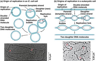
DNA replication begins at specific sites called origins of replication. These origins are recognized by replication enzymes and mark the starting point for DNA synthesis.
Bacterial Cells: Typically have a single origin per chromosome.
Eukaryotic Cells: Have hundreds or thousands of origins per chromosome.
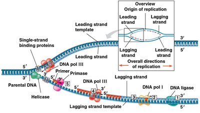
Replication Bubble: Enzymes separate the DNA strands, forming a bubble from which replication proceeds in both directions.
Replication Forks: Each bubble has two Y-shaped replication forks where DNA is unwound and new strands are synthesized.
Key Enzymes and Proteins in DNA Replication
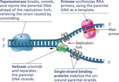
Several specialized enzymes and proteins are essential for the unwinding and stabilization of DNA during replication.
Helicase: Untwists and separates the DNA double helix at the replication forks.
Single-Strand Binding Proteins (SSBPs): Bind to and stabilize single DNA strands, preventing them from re-annealing.
Topoisomerase: Relieves strain ahead of the replication fork by breaking, swiveling, and rejoining DNA strands.
Primase: Synthesizes short RNA primers to initiate DNA synthesis.
Synthesizing a New DNA Strand
Once the DNA strands are unwound, they serve as templates for the synthesis of new complementary strands. DNA polymerases are the enzymes responsible for elongating these new strands, but they require a primer to begin synthesis.
Primer: A short stretch of RNA (5–10 nucleotides) synthesized by primase, complementary to the template strand.
DNA Polymerase III: Main enzyme for elongating new DNA strands in prokaryotes.
DNA Polymerase I: Replaces RNA primers with DNA nucleotides.
Eukaryotes: Have multiple DNA polymerases with specialized functions.
Requirements for DNA Synthesis
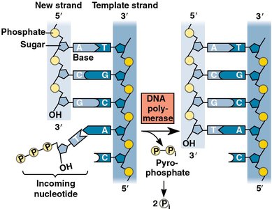
A primer with a free 3′ end
A DNA template strand
Source and Energy for Nucleotide Addition
Incoming nucleotides are nucleoside triphosphates (dATP, dTTP, dGTP, dCTP).
Energy for DNA synthesis comes from the hydrolysis of pyrophosphate (P-Pi) into two inorganic phosphates (2Pi).
Antiparallel Structure and Directionality
The two strands of DNA are antiparallel, meaning they run in opposite directions. This orientation is crucial for replication.
One strand runs 5′ → 3′, the other 3′ → 5′.
DNA polymerases can only add nucleotides to the free 3′ end, so new strands are always synthesized in the 5′ → 3′ direction.
Leading and Lagging Strands
At each replication fork, two new strands are synthesized:
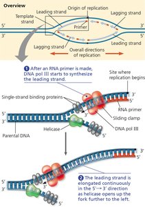
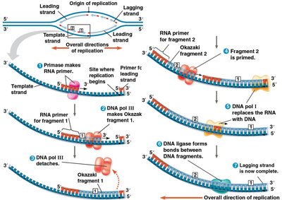
Leading Strand: Synthesized continuously in the direction of the replication fork movement. Requires only one primer.
Lagging Strand: Synthesized discontinuously away from the fork in short segments called Okazaki fragments. Each fragment requires its own primer.
Okazaki Fragments and Enzyme Roles
Okazaki Fragments: Short DNA segments synthesized on the lagging strand (1,000–2,000 nucleotides in prokaryotes; 100–200 in eukaryotes).
DNA Polymerase I: Replaces RNA primers with DNA nucleotides.
DNA Ligase: Joins the sugar-phosphate backbones of Okazaki fragments, forming a continuous strand.
Summary Table: Bacterial DNA Replication Proteins and Their Functions
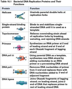
Protein | Function |
|---|---|
Helicase | Unwinds parental double helix at replication forks |
Single-strand binding protein | Binds to and stabilizes single-stranded DNA until it is used as a template |
Topoisomerase | Relieves overwinding strain ahead of replication forks by breaking, swiveling, and rejoining DNA strands |
Primase | Synthesizes an RNA primer at 5′ end of leading strand and at 5′ end of each Okazaki fragment of lagging strand |
DNA pol III | Using parental DNA as a template, synthesizes new DNA strand by adding nucleotides to an RNA primer or a pre-existing DNA strand |
DNA pol I | Removes RNA nucleotides of primer and replaces them with DNA nucleotides added to 3′ end of adjacent fragment |
DNA ligase | Joins Okazaki fragments of lagging strand; on leading strand, joins 3′ end of DNA that replaces primer to rest of leading strand DNA |
Practice Questions and Applications
Leading vs. Lagging Strands: The leading strand is synthesized continuously toward the replication fork, while the lagging strand is synthesized discontinuously away from the fork in Okazaki fragments.
Enzyme Mutations: Mutations in DNA polymerase III affect both leading and lagging strand synthesis; mutations in primase affect both strands; mutations in DNA ligase primarily affect the lagging strand.
Complementary Strand Example: For the DNA strand 5’-AACGGTCCAGTCCAAGTTACG-3’, the complementary strand is 3’-TTGCCAGGTCAGGTTCAATGC-5’.
Additional info:
DNA replication is semiconservative: each daughter DNA molecule consists of one parental strand and one newly synthesized strand.
Replication is highly regulated to ensure genome integrity and prevent mutations.
