 Indietro
DNA Structure and Replication: Foundations and Mechanisms
Guida di studio - Note intelligenti
 Appunti personalizzati basati sui tuoi materiali, ampliati con definizioni chiave, esempi e contesto.
DNA and Chromosomes
Organization of Genetic Material
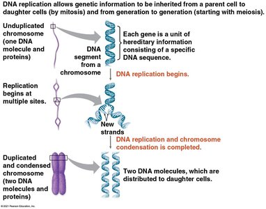
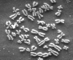
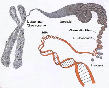
DNA is the hereditary material in almost all living organisms, organized into structures called chromosomes. Each chromosome consists of a single, long DNA molecule associated with proteins, which help package and regulate the DNA.
Chromatin: The complex of DNA and proteins (mainly histones) that forms chromosomes within the nucleus of eukaryotic cells.
Chromosome condensation: During cell division, chromatin condenses to form visible chromosomes.
Gene: A segment of DNA that encodes a functional product, usually a protein.
Structure of DNA
Nucleotide Composition and DNA Structure
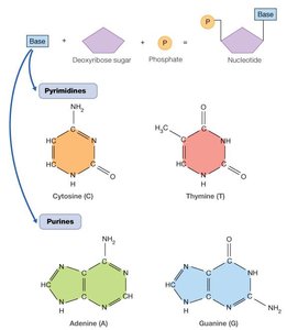
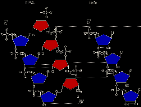
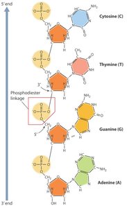
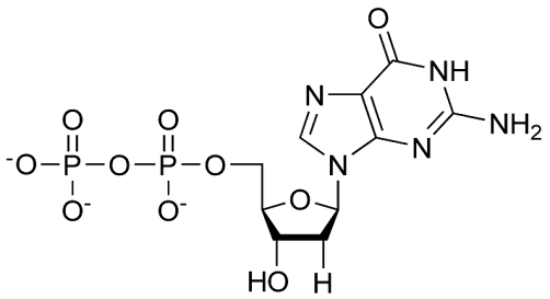
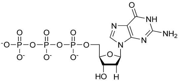
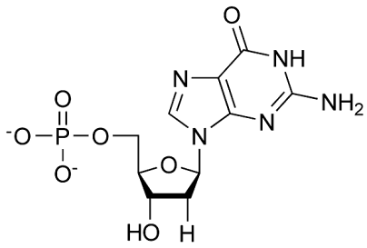
DNA is a polymer made up of repeating units called nucleotides. Each nucleotide consists of a deoxyribose sugar, a phosphate group, and a nitrogenous base. The sequence of these bases encodes genetic information.
Nitrogenous bases: Divided into purines (adenine, guanine) and pyrimidines (cytosine, thymine).
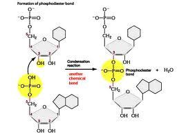
Phosphodiester bonds: Link the 5' phosphate of one nucleotide to the 3' hydroxyl of the next, forming the sugar-phosphate backbone.
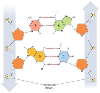
Antiparallel strands: DNA consists of two strands running in opposite directions (5' to 3' and 3' to 5').
Base pairing: Adenine pairs with thymine (A=T), and guanine pairs with cytosine (G≡C) via hydrogen bonds.
DNA as the Genetic Material
Experimental Evidence
Several key experiments established DNA as the genetic material:
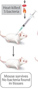
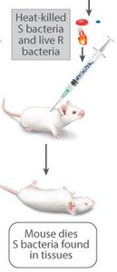
Griffith's Experiment (1928): Demonstrated transformation in Streptococcus pneumoniae—non-virulent bacteria could acquire virulence from heat-killed virulent bacteria.
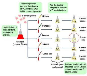
Avery, MacLeod, and McCarty (1944): Showed that DNA, not protein, was the transforming principle.
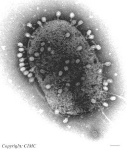
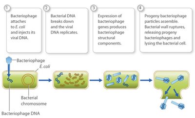
Hershey-Chase Experiment (1952): Used bacteriophages labeled with radioactive isotopes to confirm that DNA, not protein, enters bacterial cells and directs viral replication.

The Double Helix Model
Watson and Crick's Discovery
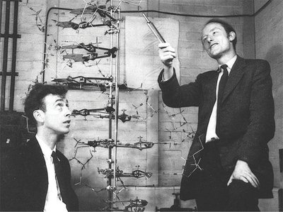
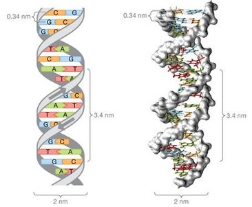
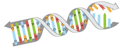
The structure of DNA was elucidated by James Watson and Francis Crick in 1953, with crucial contributions from Rosalind Franklin and Maurice Wilkins. The double helix model explained how genetic information is stored and replicated.
Antiparallel orientation: The two DNA strands run in opposite directions.
Base pairing rules: A pairs with T via two hydrogen bonds; G pairs with C via three hydrogen bonds.
Major and minor grooves: The helical structure creates grooves that are important for protein-DNA interactions.
DNA Replication
Models of Replication
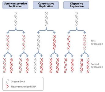
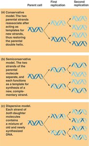
DNA replication is the process by which a cell copies its DNA before cell division. Three models were proposed: conservative, semi-conservative, and dispersive. The semi-conservative model, supported by the Meselson-Stahl experiment, is correct.
Semi-conservative replication: Each new DNA molecule consists of one parental strand and one newly synthesized strand.
Conservative replication: Parental strands reassociate after acting as templates (not supported).
Dispersive replication: Each strand is a mix of old and new DNA (not supported).
Mechanism of DNA Replication
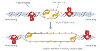
DNA replication begins at specific sites called origins of replication and proceeds bidirectionally, forming replication forks. Multiple enzymes and proteins coordinate the process.
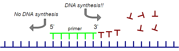
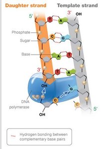
DNA polymerase: Synthesizes new DNA by adding nucleotides to a pre-existing chain (requires a primer and template).
Primase: Synthesizes a short RNA primer to provide a starting point for DNA polymerase.
Helicase: Unwinds the DNA double helix at the replication fork.
Single-stranded binding proteins (SSBPs): Stabilize unwound DNA strands.
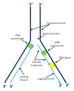
Ligase: Seals nicks between Okazaki fragments on the lagging strand.
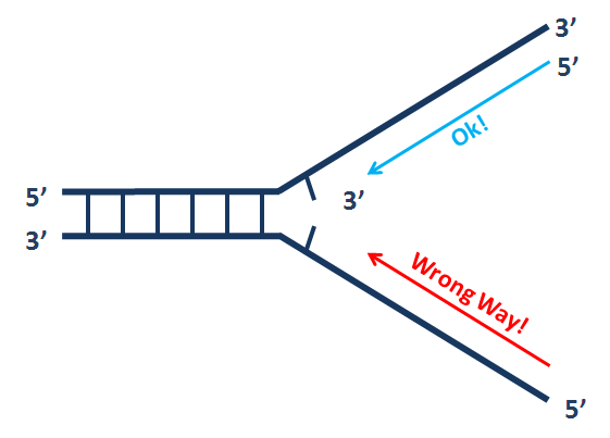
Directionality: DNA polymerase can only add nucleotides to the 3' end, so synthesis occurs 5' to 3'.
Leading and Lagging Strands
Because DNA polymerase can only synthesize in the 5' to 3' direction, replication is continuous on the leading strand and discontinuous on the lagging strand (forming Okazaki fragments).
Leading strand: Synthesized continuously toward the replication fork.
Lagging strand: Synthesized discontinuously away from the fork in short segments (Okazaki fragments), later joined by DNA ligase.
Enzymes and Proteins in DNA Replication
Key Enzymes and Their Functions
Enzyme | Function | Location |
|---|---|---|
Helicase | Unwinds the DNA double helix | Replication fork |
Ligase | Joins Okazaki fragments | Lagging strand |
DNA Polymerase I | Removes RNA primers, fills in with DNA | Both strands |
DNA Polymerase III | Main enzyme for DNA synthesis | Both strands |
Primase | Synthesizes RNA primers | Both strands |
Telomerase | Extends telomeres in eukaryotes | Chromosome ends |
Proofreading and DNA Repair
Maintaining DNA Fidelity
DNA polymerases have proofreading ability to correct errors during replication. Additional repair mechanisms fix damage or mismatches that escape proofreading.
Mismatch repair: Corrects errors missed by DNA polymerase.
Excision repair: Removes damaged DNA segments and fills the gap.
Mutations: Permanent changes in DNA sequence if errors are not corrected.
Replicating Chromosome Ends: Telomeres and Telomerase
Challenges of Linear DNA Replication
Linear chromosomes in eukaryotes pose a problem for complete replication of the ends (telomeres). Telomerase extends telomeres to prevent loss of genetic information in certain cell types.
Telomeres: Repetitive, noncoding DNA at chromosome ends.
Telomerase: Enzyme that adds telomeric repeats to chromosome ends, active in germ cells and some stem/cancer cells.
Polymerase Chain Reaction (PCR) vs. Cellular DNA Replication
Comparison Table
Feature | PCR | Cellular DNA Replication |
|---|---|---|
Primer Type | DNA | RNA |
Enzyme | Heat-stable DNA polymerase (e.g., Taq) | DNA polymerase III (prokaryotes), DNA polymerases α, δ, ε (eukaryotes) |
Template | Defined region | Entire genome/chromosome |
Initiation | Temperature cycling | Origin of replication |
Similarities: Both require primers, nucleotides, template DNA, and polymerase; both synthesize DNA 5' to 3'.
Differences: PCR uses DNA primers and amplifies specific regions; cellular replication uses RNA primers and copies the whole genome.
Key Terms and Concepts
Phosphodiester bond: Covalent bond linking nucleotides in DNA/RNA.
Hydrogen bond: Weak bond between complementary bases (A-T, G-C).
Okazaki fragments: Short DNA fragments synthesized on the lagging strand.
Semiconservative replication: Each new DNA molecule has one old and one new strand.
