 Indietro
Eukaryotic Cell Structure and Function: Mini-Textbook Study Notes
Guida di studio - Note intelligenti
 Appunti personalizzati basati sui tuoi materiali, ampliati con definizioni chiave, esempi e contesto.
Eukaryotic Cell Structure and Function
Introduction to Eukaryotic Cells
Eukaryotic cells are distinguished from prokaryotic cells by their complex internal structure, including a nucleus and various membrane-bound organelles. These features allow eukaryotes to carry out specialized functions and support larger cell sizes.
Nucleus: Contains DNA enclosed by a nuclear envelope.
Membrane-bound organelles: Specialized compartments for distinct cellular processes.
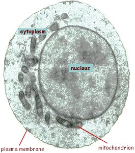
Cytoplasm: Region between the plasma membrane and nucleus.
Cell size: Eukaryotic cells are generally larger than prokaryotic cells.
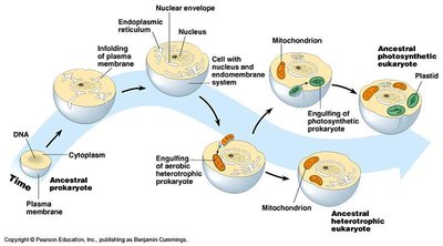
The Nucleus
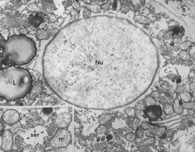
The nucleus is the control center of the cell, housing genetic material and coordinating activities such as growth and reproduction.
Nuclear envelope: Double membrane structure with nuclear pores for molecular transport.
Nuclear pores: Complexes that regulate entry and exit of proteins and RNA.
Chromatin: DNA-protein complex associated with the inner membrane.
Nucleolus: Site of ribosomal RNA synthesis.
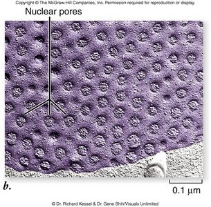
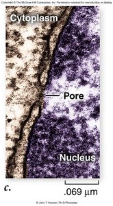
Structure and Function of the Nuclear Envelope
The nuclear envelope is perforated by nuclear pore complexes, which are essential for the selective transport of molecules.
Nuclear localization signal (NLS): A 17-amino-acid sequence that directs nuclear proteins to the nucleus.
Eukaryotic Ribosomes
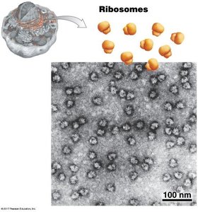
Ribosomes are universal machines for protein synthesis, found in all domains of life. Eukaryotic ribosomes are composed of two subunits, each containing rRNA and protein, and are located in various cellular compartments.
Subunit composition: Two subunits (large and small), each with rRNA and protein.
Location: Free in cytoplasm or attached to the endoplasmic reticulum.
Function: Protein synthesis for different cellular destinations.
The Endoplasmic Reticulum (ER)
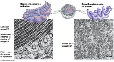
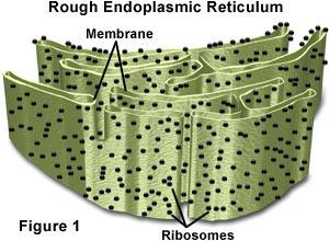
The ER is an organelle continuous with the nuclear envelope, forming a network of tubules and sacs (cisternae). It is divided into two main types:
Rough ER (RER): Studded with ribosomes; synthesizes secreted and membrane proteins.
Smooth ER (SER): Lacks ribosomes; synthesizes lipids and detoxifies chemicals.
Ratio of RER/SER: Varies depending on cell function (e.g., lipid synthesis in testes, protein secretion in immune cells).
The Golgi Apparatus
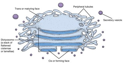
The Golgi apparatus is a membranous organelle composed of stacks of flattened cisternae (dictyosomes). It is present in most eukaryotic cells and is essential for modifying, sorting, and packaging proteins and lipids.
Structure: Stacks of cisternae with distinct cis (forming) and trans (maturing) faces.
Function: Processing and trafficking of proteins and lipids.
Movement of Materials: The Endomembrane System
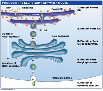
The endomembrane system includes the ER, Golgi apparatus, lysosomes, and vesicles, coordinating the synthesis, modification, and transport of proteins and lipids.
Protein targeting: Most organelle proteins are imported from the cytosol via signal sequences.
Lysosomes
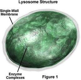
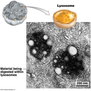
Lysosomes are recycling centers found only in animal cells. They contain acid hydrolases that degrade macromolecules at an acidic pH maintained by proton pumps.
Enzymes: Acid hydrolases function optimally at pH 5.0.
Function: Breakdown and recycling of cellular waste.
Vacuoles
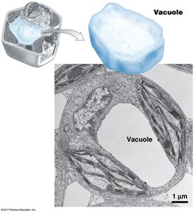
Vacuoles are large, membrane-bound structures found in plants and fungi. They serve various functions, including digestion and storage of water and ions to maintain cell volume.
Specialization: Some vacuoles are specialized for digestion; most store water and ions.
Mitochondria
Mitochondria are the "powerhouses" of the cell, generating ATP through electron transport and oxidative phosphorylation. They are found in all eukaryotic cells and may exist in large numbers.
ATP production: Via electron transport chain and oxidative phosphorylation.
Tricarboxylic acid cycle: Central metabolic pathway for energy production.
Size: Comparable to bacteria.
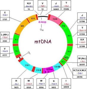
mtDNA: Mitochondria contain their own DNA and can reproduce independently.
Chloroplasts
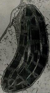
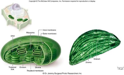
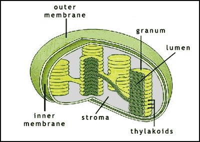
Chloroplasts are organelles found in plants and algae, responsible for photosynthesis. They are surrounded by two membranes and contain a stroma, thylakoids, and grana.
Stroma: Gelatinous matrix containing DNA, ribosomes, and other components.
Thylakoids: Flattened sacs where light reactions occur.
Grana: Stacks of thylakoids.
Photosynthesis: Light reactions generate ATP, NADPH, and O2; dark reactions form carbohydrates.
Cytoplasm and Cytoplasmic Matrix
The cytoplasm is a complex, differentiated matrix within the cell membrane, providing an environment for cellular activities.
Composition: 70-85% water by volume; half is free water, half is bound to macromolecules.
pH: Ranges from 6.8-7.1; vacuoles may reach pH ~3.
Structure: Often semi-crystalline due to high protein content.
Cytoskeleton: Microfilaments provide shape and movement.
The Cytoskeleton
The cytoskeleton is a dynamic network of microtubules, intermediate filaments, and microfilaments, essential for cell shape, movement, and organelle positioning.
Microtubules: 25 nm diameter, composed of α and β tubulin.
Intermediate filaments: 8-10 nm diameter.
Microfilaments: 4-7 nm diameter, composed of actin.
Function: Cell shape, movement, anchoring, and transport of organelles.
Prokaryotes: Lack a true, organized cytoskeleton.
External Cell Coverings
Not all eukaryotes have cell walls. Plants, fungi, and some protists do, with variable composition.
Algae/plants: Cellulose and pectin.
Diatoms: Silica.
Fungi: Chitin, cellulose, and glycan.
Cilia and Flagella
Cilia and flagella are prominent organelles associated with movement. They differ in size and movement patterns but share a similar ultrastructure.
Cilia: 5-20 μm long; beat with effective and recovery phases.
Flagella: 100-200 μm long; move in an undulating fashion.
Structure: Membrane-bound cylinders (~2 μm diameter) with an axoneme (9+2 microtubule arrangement).
Molecular Unity of Prokaryotes and Eukaryotes
Despite structural differences, prokaryotes and eukaryotes share fundamental molecular features.
Chemical composition: Similar basic molecules.
Genetic code: Universal among all life forms.
Metabolic processes: Shared pathways and origins.
Summary Table: Major Eukaryotic Organelles and Their Functions
Organelle | Main Function | Key Features |
|---|---|---|
Nucleus | Genetic information storage, transcription | Double membrane, nuclear pores, nucleolus |
Ribosomes | Protein synthesis | Two subunits, rRNA and protein |
ER (Rough/Smooth) | Protein and lipid synthesis | Rough: ribosomes; Smooth: lipid synthesis |
Golgi Apparatus | Protein modification and sorting | Stacks of cisternae, cis/trans faces |
Lysosomes | Macromolecule degradation | Acid hydrolases, low pH |
Vacuoles | Storage, digestion | Large, membrane-bound |
Mitochondria | ATP production | Double membrane, mtDNA |
Chloroplasts | Photosynthesis | Double membrane, thylakoids, grana |
Cytoskeleton | Shape, movement | Microtubules, filaments |
Key Equations
ATP Production (Oxidative Phosphorylation):
Photosynthesis (Overall Reaction):
Additional info:
Some details were inferred for completeness, such as the universal nature of the genetic code and the basic metabolic processes shared by prokaryotes and eukaryotes. The summary table was constructed to provide a concise comparison of organelle functions.
