 Indietro
General Biology Study Guide: Cell Signaling, Chromosome Structure, and Cell Cycle
Guida di studio - Note intelligenti
 Appunti personalizzati basati sui tuoi materiali, ampliati con definizioni chiave, esempi e contesto.
Q1. Label the diagram showing the movement of molecules across the plasma membrane.
Background
Topic: Cell Membrane Transport
This question tests your understanding of how molecules move between the extracellular fluid and the cytoplasm, including passive and active transport mechanisms.
Key Terms:
Plasma membrane
Extracellular fluid
Cytoplasm
Diffusion
Active transport
Step-by-Step Guidance
Examine the diagram and identify the direction of molecule movement (from extracellular fluid to cytoplasm or vice versa).
Determine if the movement is passive (diffusion) or requires energy (active transport).
Label the boxes with the appropriate transport mechanism based on the direction and type of movement.
Consider the role of the plasma membrane in regulating what enters and exits the cell.


Try solving on your own before revealing the answer!
Final Answer:
The boxes should be labeled as follows: left box - "Passive transport (diffusion)", middle box - "Active transport", right box - "Facilitated diffusion". The plasma membrane regulates these processes.
Q2. Sequence the steps in the cell signaling pathway shown in the diagram.
Background
Topic: Cell Signaling
This question tests your understanding of how a signal is received at the cell surface and transduced through a series of molecular events inside the cell.
Key Terms:
Signal transduction
Receptor
Second messenger
Effector protein
Step-by-Step Guidance
Identify the initial signal molecule and the receptor it binds to on the cell membrane.
Follow the arrows to see how the signal is relayed through intermediate molecules.
Determine which molecules act as second messengers and which are effector proteins.
Label each step in the pathway according to its function (signal reception, transduction, response).

Try solving on your own before revealing the answer!
Final Answer:
The steps are: signal molecule binds receptor, receptor activates relay proteins, relay proteins activate second messengers, second messengers activate effector proteins, leading to a cellular response.
Q3. Label the parts of the chromosome structure diagram.
Background
Topic: Chromosome Structure
This question tests your knowledge of the anatomy of a chromosome, including centromeres, chromatids, and telomeres.
Key Terms:
Chromatid
Centromere
Telomere
Chromosome
Step-by-Step Guidance
Examine the diagram and locate the central constriction (centromere).
Identify the arms of the chromosome (chromatids).
Find the ends of the chromosome (telomeres).
Label each part according to its function and location.




Try solving on your own before revealing the answer!
Final Answer:
The labels are: centromere (central constriction), chromatids (arms), telomeres (ends), chromosome (entire structure).
Q4. Identify the stages and key structures in the cell cycle diagram.
Background
Topic: Cell Cycle
This question tests your understanding of the phases of the cell cycle and the checkpoints that regulate cell division.
Key Terms:
G1 phase
S phase
G2 phase
M phase
Checkpoint
Cyclin
Step-by-Step Guidance
Identify the different phases of the cell cycle in the diagram (G1, S, G2, M).
Locate the checkpoints and label them.
Find the cyclin and CDK complexes and their roles in cell cycle regulation.
Label each structure according to its function in the cell cycle.




Try solving on your own before revealing the answer!
Final Answer:
The stages are G1, S, G2, and M. Key structures include checkpoints (G1, G2, M), cyclin, and CDK complexes that regulate progression through the cycle.
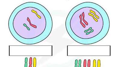
Q5. Compare the chromosome arrangements in the two cells shown and identify the process illustrated.
Background
Topic: Meiosis and Genetic Variation
This question tests your understanding of how chromosomes are arranged during meiosis and how genetic variation is introduced.
Key Terms:
Homologous chromosomes
Meiosis
Genetic recombination
Independent assortment
Step-by-Step Guidance
Examine the arrangement of chromosomes in each cell.
Identify whether the chromosomes are homologous pairs or sister chromatids.
Determine which stage of meiosis is illustrated.
Label the process and explain how it contributes to genetic variation.
Try solving on your own before revealing the answer!
Final Answer:
The diagram shows homologous chromosomes arranged differently in two cells, illustrating independent assortment during meiosis, which increases genetic variation.
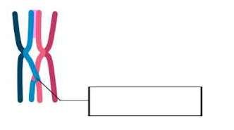
Q6. Identify the process shown in the chromosome crossover diagram and explain its significance.
Background
Topic: Genetic Recombination
This question tests your understanding of crossing over during meiosis and its role in genetic diversity.
Key Terms:
Crossing over
Chiasma
Genetic recombination
Meiosis
Step-by-Step Guidance
Examine the diagram and identify the point where two chromosomes overlap.
Label the chiasma, where crossing over occurs.
Explain how crossing over leads to genetic recombination.
Describe the significance of this process in meiosis.
Try solving on your own before revealing the answer!
Final Answer:
The process is crossing over at the chiasma, which allows for genetic recombination and increases genetic diversity during meiosis.
