 Indietro
General Biology Study Guide: Cell Structure, Membrane Function, Metabolism, and Cellular Respiration
Guida di studio - Note intelligenti
 Appunti personalizzati basati sui tuoi materiali, ampliati con definizioni chiave, esempi e contesto.
Chapter 6 - A Tour of the Cell
Prokaryotic vs. Eukaryotic Cells
Cells are classified into two main types: prokaryotic and eukaryotic. These classifications are based on structural and functional differences.
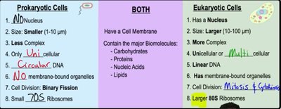
Prokaryotic Cells: Do not have a nucleus, are smaller (1-10 μm), less complex, usually unicellular, possess circular DNA, lack membrane-bound organelles, divide by binary fission, and contain small 70S ribosomes.
Eukaryotic Cells: Have a nucleus, are larger (10-100 μm), more complex, can be unicellular or multicellular, possess linear DNA, contain membrane-bound organelles, divide by mitosis and cytokinesis, and have larger 80S ribosomes.
Both: Have a cell membrane and contain major biomolecules such as carbohydrates, proteins, nucleic acids, and lipids.
Example: Bacteria are prokaryotic, while plants and animals are eukaryotic.
Animal vs. Plant Cells
Animal and plant cells are both eukaryotic but differ in certain organelles and structures.
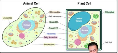
Animal Cells: Contain lysosomes, mitochondria, cell membrane, rough and smooth ER, ribosomes, Golgi apparatus, and peroxisomes.
Plant Cells: Contain a central vacuole, chloroplasts, cell wall, mitochondria, cell membrane, rough and smooth ER, ribosomes, Golgi apparatus, and peroxisomes.
Key Differences: Plant cells have a cell wall, chloroplasts, and a central vacuole, while animal cells have lysosomes.
Example: Chloroplasts are found only in plant cells and are responsible for photosynthesis.
Microscope Types
Microscopes are essential tools for visualizing cells and their structures.
Light Microscopes: Use light to magnify small objects.
Electron Microscopes: Use electrons for higher magnification. Includes Scanning Electron Microscope (SEM) for external surfaces and Transmission Electron Microscope (TEM) for internal structures.
Example: TEM is used to study the internal structure of mitochondria.
Eukaryotic Organelles
Eukaryotic cells contain specialized organelles, each with distinct functions.
Nucleus: Stores DNA and controls cellular activities. Surrounded by a nuclear envelope with pores; contains the nucleolus for ribosome assembly.
Ribosomes: Build proteins via translation; can be free or attached to rough ER.
Endoplasmic Reticulum (ER): Rough ER modifies proteins; smooth ER synthesizes lipids and detoxifies substances.
Golgi Apparatus: Processes and packages proteins and lipids for secretion.
Lysosomes: Digest and recycle cellular waste; found mainly in animal cells.
Central Vacuole: Stores water and ions; maintains turgor pressure in plant cells.
Mitochondria: Site of cellular respiration; produces ATP.
Chloroplasts: Site of photosynthesis in plant cells; contains thylakoids and stroma.
Peroxisomes: Conduct oxidation reactions and detoxify harmful substances.
Cytoskeleton: Provides structure, movement, and transport; includes microfilaments, intermediate filaments, and microtubules.
Cell Wall: Provides protection and structure in plant cells; made of cellulose.
Extracellular Matrix (ECM): Found in animal cells; composed of glycoproteins for cell anchoring.
Cell Junctions: Connect adjacent cells; types include tight junctions and desmosomes.
Example: Mitochondria are known as the "powerhouse of the cell" due to their role in ATP production.
Chapter 7 - Membrane Structure and Function
Biological Membranes
Biological membranes are primarily composed of phospholipids and proteins, forming a fluid mosaic structure.
Phospholipids: Amphipathic molecules with hydrophilic heads and hydrophobic tails.
Fluid Mosaic Model: Membranes are dynamic, with proteins moving laterally within the lipid bilayer.
Membrane Proteins: Functions include recognition, anchorage, signal transduction, transport, linkage, and enzymatic activity.
Example: Transport proteins facilitate the movement of ions across the membrane.
Concentration Gradients and Diffusion
Substances move across membranes according to concentration gradients.
Diffusion: Movement from high to low concentration; no energy required.
Active Transport: Movement from low to high concentration; requires energy.
Example: Oxygen diffuses into cells down its concentration gradient.
Membrane Transport
Membranes are selectively permeable, allowing only certain molecules to cross freely.
Freely Diffusing Molecules: Small, uncharged, nonpolar/hydrophobic.
Restricted Molecules: Large, charged, polar/hydrophilic.
Example: Water crosses membranes via aquaporins (facilitated diffusion).
Passive vs. Active Transport
Transport across membranes can be passive or active.
Passive Transport: No energy required; includes simple and facilitated diffusion.
Active Transport: Requires energy; includes primary (ATP-driven) and secondary (gradient-driven) transport.
Example: The Na+/K+ pump is a primary active transport mechanism.
Osmosis and Tonicity
Osmosis is the passive diffusion of water across membranes, influenced by solute concentration (tonicity).
Hypotonic: Lower solute concentration; water enters cells.
Isotonic: Equal solute concentration; water movement is balanced.
Hypertonic: Higher solute concentration; water exits cells.
Example: Plant cells prefer hypotonic environments for turgor pressure.
Endocytosis and Exocytosis
Large molecules are transported via vesicle-mediated processes.
Endocytosis: Entry of macromolecules via vesicle formation; includes phagocytosis, pinocytosis, and receptor-mediated endocytosis.
Exocytosis: Exit of molecules via vesicle fusion with the membrane.
Example: Hormones are released from cells by exocytosis.
Chapter 8 - An Introduction to Metabolism
Energy and Thermodynamics
Energy is the ability to perform work, and biological systems obey the laws of thermodynamics.
Potential Energy: Stored energy (e.g., chemical bonds).
Kinetic Energy: Energy of motion (e.g., muscle contractions).
First Law: Energy cannot be created or destroyed; total energy remains constant.
Second Law: Energy conversions increase entropy (disorder); heat is lost.
Example: Cellular respiration transforms chemical energy into ATP and heat.
Chemical Reactions
Chemical reactions involve making or breaking bonds, leading to changes in matter.
Endergonic: Require energy input.
Exergonic: Release energy.
Example: ATP hydrolysis is an exergonic reaction.
ATP and Energy Coupling
ATP is the primary energy carrier in cells, coupling exergonic and endergonic reactions.
ATP Structure: Three phosphate groups, pentose sugar, adenine base.
ATP Hydrolysis: Releases energy by breaking phosphate bonds.
Phosphorylation: Transfer of phosphate group to another molecule, activating it.
Example: ATP powers muscle contraction by phosphorylating proteins.
Enzymes and Catalysis
Enzymes are biological catalysts that speed up reactions by lowering activation energy.
Substrate: Reactant in an enzyme-catalyzed reaction.
Active Site: Region where substrate binds.
Enzyme-Substrate Complex: Temporary association during catalysis.
Cofactors: Non-protein helpers; coenzymes are organic cofactors.
Inhibitors: Competitive (bind active site) or noncompetitive (bind allosteric site).
Example: Digestive enzymes break down food in the stomach.
Metabolic Pathways
Metabolism encompasses all chemical reactions in an organism, organized into pathways.
Catabolic Pathways: Break down molecules, releasing energy.
Anabolic Pathways: Build up molecules, requiring energy.
Feedback Regulation: Negative feedback inhibits pathways; positive feedback stimulates them.
Example: Glycolysis is a catabolic pathway; protein synthesis is anabolic.
Chapter 9 - Cellular Respiration and Fermentation
Redox Reactions
Cellular respiration involves redox reactions, transferring electrons between molecules.
Oxidation: Loss of electrons.
Reduction: Gain of electrons.
Electron Carriers: NADH and FADH2 transport electrons.
Example: NADH carries electrons to the electron transport chain.
Stages of Cellular Respiration
Aerobic cellular respiration consists of four main stages:
Glycolysis: Breaks down glucose into 2 pyruvate; occurs in cytoplasm; produces 2 ATP and 2 NADH.
Pyruvate Oxidation: Converts pyruvate to acetyl-CoA; produces NADH and CO2.
Krebs Cycle: Oxidizes acetyl-CoA; produces ATP, NADH, FADH2, and CO2.
Electron Transport Chain & Chemiosmosis: Uses NADH and FADH2 to generate ATP via oxidative phosphorylation.
Example: The ETC uses oxygen as the final electron acceptor, forming water.
Types of Phosphorylation
Substrate-Level Phosphorylation: Direct transfer of phosphate to ADP by an enzyme.
Oxidative Phosphorylation: Uses energy from electron transport and chemiosmosis to make ATP.
Example: Most ATP is produced by oxidative phosphorylation in mitochondria.
Fermentation and Anaerobic Respiration
When oxygen is unavailable, cells use fermentation or anaerobic respiration to generate ATP.
Fermentation: Regenerates NAD+ by reducing pyruvate; produces lactic acid or ethanol.
Anaerobic Respiration: Uses alternative electron acceptors (e.g., nitrate, sulfate); produces more ATP than fermentation but less than aerobic respiration.
Example: Yeast produces ethanol during alcohol fermentation.
Summary Table: Products of Cellular Respiration
Stage | Start Molecule | CO2 | ATP | FADH2 | NADH | End Molecule |
|---|---|---|---|---|---|---|
Glycolysis | Glucose | 0 | 2 | 0 | 2 | 2 Pyruvate |
Pyruvate Oxidation | 2 Pyruvate | 2 | 0 | 0 | 2 | 2 Acetyl-CoA |
Krebs Cycle | 2 Acetyl-CoA | 2 | 2 | 2 | 6 | Oxaloacetate |
Oxidative Phosphorylation | NADH and FADH2 | 0 | 26-34 | 0 | 0 | H2O |
Total | 6 | 30-38 | 2 | 10 |
Additional info: The table summarizes the main products of each stage of aerobic cellular respiration, including ATP yield and electron carriers.
