 Indietro
Hypersensitivity Reactions: Types, Mechanisms, and Clinical Examples
Guida di studio - Note intelligenti
 Appunti personalizzati basati sui tuoi materiali, ampliati con definizioni chiave, esempi e contesto.
Hypersensitivity Reactions
Overview of Hypersensitivity
Hypersensitivity refers to excessive or inappropriate immune responses that result in host tissue damage following prolonged or repeated exposure to antigens. These reactions are classified based on their immune mediators, response time, and mechanisms of activation. Understanding hypersensitivity is crucial for recognizing allergic diseases and autoimmune disorders.
Definition: Hypersensitivity is an exaggerated immune response causing tissue injury.
Classification: Four main types: Type I (Immediate), Type II (Cytotoxic), Type III (Immune Complex), Type IV (Cell-mediated).
Key Symptoms: Inflammation, edema, tissue damage, and organ dysfunction.

Classification of Hypersensitivity Reactions
Each type of hypersensitivity is mediated by distinct immune components and has characteristic clinical manifestations.
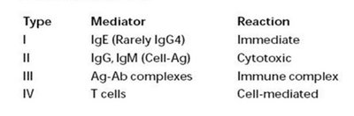
Type | Mediator | Reaction |
|---|---|---|
I | IgE (Rarely IgG4) | Immediate |
II | IgG, IgM (Cell-Ag) | Cytotoxic |
III | Ag-Ab complexes | Immune complex |
IV | T cells | Cell-mediated |
Type I Hypersensitivity (Immediate/Allergic)
Mechanism and Pathogenesis
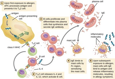
Type I hypersensitivity is the most common form of allergic reaction, characterized by rapid onset (minutes after exposure). It is mediated by IgE antibodies, which trigger mast cell and basophil degranulation, releasing inflammatory mediators.
Initial Exposure: Allergen is recognized by B-lymphocytes, which differentiate into plasma cells producing IgE.
Sensitization: IgE binds to Fc receptors on mast cells.
Re-exposure: Allergen cross-links IgE on mast cells, causing degranulation and release of histamine, leukotrienes, and prostaglandins.
Immediate Phase: Vasodilation, increased vascular permeability, smooth muscle contraction.
Late Phase: Recruitment of eosinophils, neutrophils, and lymphocytes, causing further tissue damage and symptoms.
Clinical Manifestations
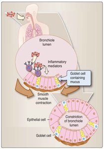
Asthma: Airway obstruction, bronchoconstriction, inflammation, and mucus production.
Allergic Rhinitis (Hay Fever): Nasal congestion, sneezing, itching, discharge, and conjunctivitis.
Allergic Eczema (Atopic Dermatitis): Itching, red patches, edema in epidermis.
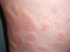
Urticaria (Hives): Itchy wheals, localized vasodilation, and edema.
Anaphylaxis: Life-threatening systemic reaction with airway constriction, hypotension, and shock.
Management of Type I Hypersensitivity
Active Avoidance: Identifying and avoiding allergens (diagnostic tests, skin tests).
Pharmacotherapy: Adrenaline for anaphylaxis; antihistamines, corticosteroids, antileukotrienes for mild reactions.
Desensitization Therapy: Gradual exposure to increasing doses of allergen to reduce sensitivity.
Type II Hypersensitivity (Cytotoxic)
Mechanism and Pathogenesis
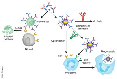
Type II hypersensitivity involves IgG or IgM antibodies directed against cell surface or extracellular matrix antigens, leading to cell destruction or dysfunction. It can be autoimmune or triggered by exogenous substances.
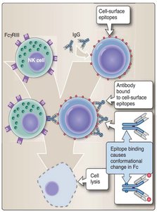
Antibody-Dependent Cell-Mediated Cytotoxicity (ADCC): Fc receptor-bearing cells (NK cells, monocytes, neutrophils, eosinophils) bind to antibody-coated cells, causing cytolysis.
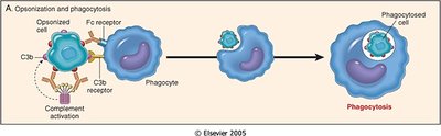
Opsonization and Phagocytosis: Antibodies and complement components act as opsonins, enhancing phagocytosis by neutrophils and macrophages.
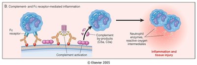
Complement- and Fc Receptor-Mediated Inflammation: Antibody binding activates complement, recruiting inflammatory cells and causing tissue injury.
Antibody-Mediated Cellular Dysfunction: Antibodies block cell surface receptors, disrupting normal cell function.

Clinical Examples
Autoimmune: Hemolytic anemia, thrombocytopenic purpura, rheumatic heart disease, Goodpasture's nephritis, Graves’ disease, myasthenia gravis.
Non-autoimmune: Transfusion reactions (ABO incompatibility), hemolytic disease of the newborn (erythroblastosis fetalis).

Type III Hypersensitivity (Immune Complex)
Mechanism and Pathogenesis
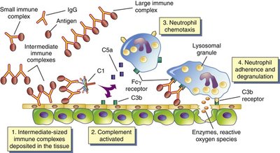
Type III hypersensitivity is caused by the formation of soluble antigen-antibody complexes that deposit in tissues, triggering inflammation and tissue damage. The reaction can be local or systemic.
Immune Complex Formation: Antibodies bind to antigens, forming complexes.
Deposition: Complexes lodge in capillaries and basement membranes, especially in skin, joints, and kidneys.
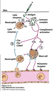
Inflammation: Complexes activate complement, recruit neutrophils, and cause mast cell degranulation, leading to tissue injury.

Clinical Manifestations
Localized Reaction (Arthus Reaction): Local tissue damage, edema, and erythema after intradermal antigen injection.
Systemic Reaction (Serum Sickness): Disseminated immune complexes cause fever, rash, vasculitis, arthritis, glomerulonephritis, and hepatitis. Examples include systemic lupus erythematosus (SLE) and rheumatoid arthritis.
Type IV Hypersensitivity (Delayed-Type/Cell-Mediated)
Mechanism and Pathogenesis
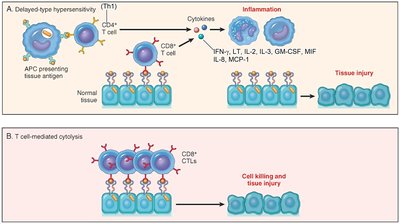
Type IV hypersensitivity is mediated by T cells and is antibody-independent. It develops over days and involves overstimulation of T cells and monocytes/macrophages, leading to cytokine release, inflammation, and tissue damage.
Delayed-Type Hypersensitivity (DTH): CD4+ T cells are activated by antigen-presenting cells, proliferate, and upon re-exposure, release cytokines that recruit macrophages and cause tissue injury.
Cell-Mediated Cytotoxicity: CD8+ T cells (CTLs) target and kill cells presenting antigen via MHC I, causing apoptosis and cytolysis.
Clinical Examples
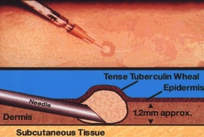
Tuberculin Hypersensitivity (Mantoux Test): Used to detect exposure to Mycobacterium tuberculosis.
Contact Dermatitis: Caused by poison ivy, latex, heavy metals.
Granulomatous Diseases: Leprosy, tuberculosis, Crohn’s disease.
Transplant Rejection: T cell-mediated destruction of transplanted tissues.
Summary Table: Types of Hypersensitivity
Type | Immune Mediator | Response Time | Mechanism | Examples |
|---|---|---|---|---|
I | IgE | Minutes | Allergen-induced mast cell degranulation | Asthma, hay fever, anaphylaxis |
II | IgG, IgM | Hours | Antibody binding to cell surface antigens | Hemolytic anemia, transfusion reaction |
III | Immune complexes | Hours to days | Immune complex deposition and inflammation | SLE, rheumatoid arthritis, Arthus reaction |
IV | T cells | Days | T cell-mediated inflammation and cytotoxicity | Tuberculin reaction, contact dermatitis |
Key Point: Allergies are adaptive immune responses to allergens, sometimes life-threatening, and are classified based on the underlying immunological mechanism.
