 Indietro
Inside the Cell: Structure and Function of Cellular Components
Guida di studio - Note intelligenti
 Appunti personalizzati basati sui tuoi materiali, ampliati con definizioni chiave, esempi e contesto.
Introduction to Microscopes
Understanding the Need for Microscopy
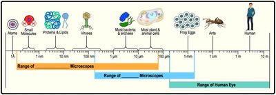
Many biological structures, such as cells and their components, are too small to be seen with the naked eye. Microscopes are essential tools that allow scientists to visualize and study these microscopic entities.
Microscope: An optical instrument used to magnify and resolve details of objects too small for the unaided eye, such as cells.
There are two main types of microscopes commonly used in biology:
Light Microscopes: Use visible light to magnify objects, suitable for viewing most cells and some organelles.
Electron Microscopes: Use beams of electrons for much higher magnification and resolution, allowing visualization of smaller structures like viruses and macromolecules.
Types of Electron Microscopes
Scanning Electron Microscope (SEM): Used to visualize the external surfaces of cells and other structures in high detail.
Transmission Electron Microscope (TEM): Used to visualize internal cell structures by passing electrons through thin sections of specimens.
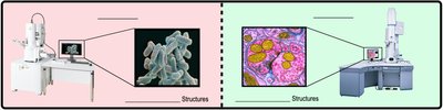
Example: To measure the size of a ribosome inside a eukaryotic cell, a transmission electron microscope (TEM) would be used.
Prokaryotic and Eukaryotic Cells
Major Cell Types
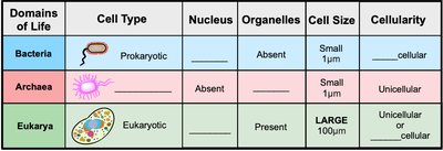
All life is classified into two broad categories based on cellular structure: prokaryotic and eukaryotic cells.
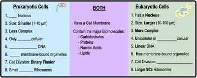
Prokaryotic Cells: Lack a nucleus and membrane-bound organelles. Includes Bacteria and Archaea.
Eukaryotic Cells: Possess a nucleus and various membrane-bound organelles. Includes Protists, Fungi, Plants, and Animals.
Features of Bacterial Cells
Bacteria are the most abundant and diverse organisms on Earth.
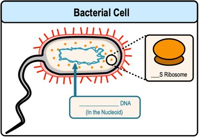
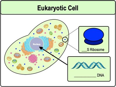
Bacterial DNA is circular and found in a region called the nucleoid.
Bacteria have small (70S) ribosomes and divide by binary fission.
Features of Eukaryotic Cells
Eukaryotic cells contain several membrane-bound organelles, including a nucleus.
Eukaryotic DNA is linear and found inside the nucleus.
Eukaryotes have large (80S) ribosomes and divide by mitosis and cytokinesis.
Comparison: Prokaryotic vs. Eukaryotic Cells
Key differences include the presence of a nucleus, organelles, cell size, and complexity.
Introduction to Eukaryotic Organelles
Overview of Organelles
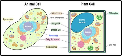
Eukaryotic cells contain a variety of organelles, each with specialized functions. Some organelles are unique to animal or plant cells.
Ribosomes
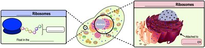
Ribosomes are sometimes called "non-membranous organelles."
They are molecular machines that build proteins in all living cells through the process of translation.
Ribosomes can be free in the cytoplasm or attached to the rough endoplasmic reticulum (ER).
Map of Eukaryotic Organelles
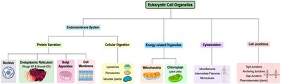
The major organelles can be grouped by their roles in protein secretion, cellular digestion, energy production, cytoskeleton, and cell junctions.
Mitochondria and Chloroplasts
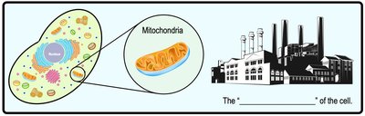
Mitochondria: The Powerhouse of the Cell
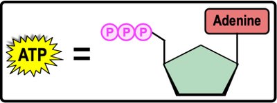
Mitochondria synthesize large amounts of ATP (adenosine triphosphate), the cell's main energy currency.
Cellular respiration occurs in mitochondria, breaking down sugars and lipids to produce ATP.
Mitochondria Structure
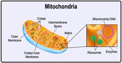
Mitochondria have their own ribosomes and DNA, independent of nuclear DNA.
They possess two membranes: an outer membrane and a highly folded inner membrane (cristae).
The intermembrane space lies between the two membranes, while the matrix is the innermost region containing enzymes, ribosomes, and mitochondrial DNA.
Chloroplasts: The Site of Photosynthesis
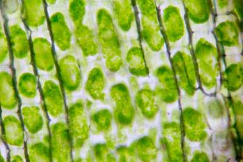
Chloroplasts are green organelles found in plant cells, responsible for photosynthesis.
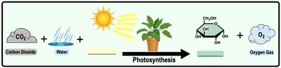
Photosynthesis uses energy from sunlight to synthesize sugars (glucose) from carbon dioxide and water, releasing oxygen as a byproduct.
Chloroplast Structure
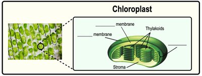
Chloroplasts have two membranes (outer and inner) without folds.
Thylakoids are interconnected disc-shaped sacs; grana are stacks of thylakoids.
The stroma is the innermost region containing enzymes, ribosomes, and chloroplast DNA.
The Endomembrane System: Protein Secretion and Digestion
Overview of the Endomembrane System
The endomembrane system is a group of membrane-bound organelles in eukaryotic cells that work together for protein secretion, molecule transport, and cellular digestion.
Organelles are interconnected by vesicles (small membrane bubbles).
Functions include protein synthesis, modification, transport, and cellular digestion.
Protein Secretion Pathway
Protein secretion involves several organelles in a specific sequence:
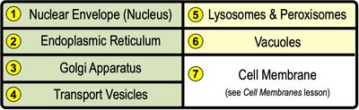
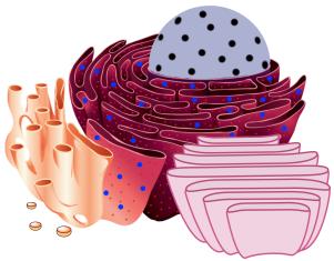
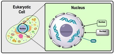
Nucleus: Stores DNA, the genetic code for proteins. The nuclear envelope surrounds the nucleus, with nuclear pores allowing entry and exit. The nucleolus assembles ribosomes.
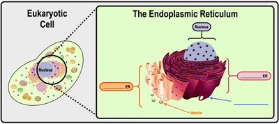
Endoplasmic Reticulum (ER): The rough ER (rER) is studded with ribosomes and synthesizes proteins, while the smooth ER (sER) synthesizes lipids and detoxifies substances.
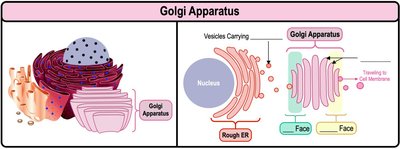
Golgi Apparatus: Receives, modifies, and repackages proteins and lipids for export.
Vesicles: Transport proteins between organelles and to the cell membrane for secretion.
Digestive Organelles: Lysosomes, Peroxisomes, and Vacuoles
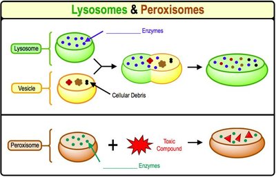
Lysosomes: Acidic vesicles containing digestive enzymes that break down food, debris, and pathogens. Primarily found in animal cells and originate from the Golgi apparatus.
Peroxisomes: Vesicles containing enzymes that break down toxic compounds (e.g., hydrogen peroxide) and fatty acids. Found in all eukaryotic cells and originate from the rough ER.
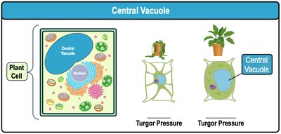
Central Vacuole: Large vesicle in plant cells that degrades and recycles molecules, and maintains turgor pressure for structural support.
The Cytoskeleton and Cell Movement
Cytoskeleton Structure and Function
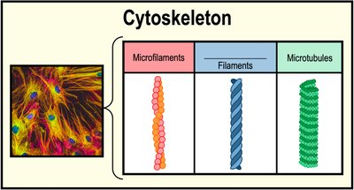
The cytoskeleton is a network of protein filaments that provides cell shape, structure, movement, intracellular transport, and signaling.
Three major components:
Microfilaments: Smallest, made of actin proteins, involved in cell movement and shape.
Intermediate Filaments: Medium-sized, made of various proteins, provide mechanical support.
Microtubules: Largest, made of tubulin proteins, form tubes for transport and cell division.
Cilia and Flagella
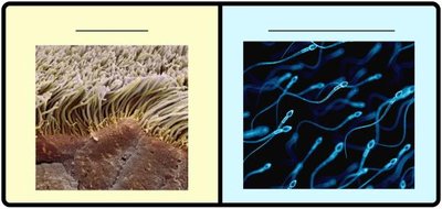
Microtubules are the main structural component of cilia and flagella, which provide cell movement.
Cilia: Multiple short, hair-like structures that move in a coordinated fashion to move objects or the cell itself.
Flagella: Longer, tail-like structures that move in a whip-like manner to propel cells.
Additional info: The cytoskeleton also plays a role in cell division, intracellular transport, and maintaining cell integrity under stress.
