 Indietro
Lipids, Membranes, and Cell Compartments: Study Guide
Guida di studio - Note intelligenti
 Appunti personalizzati basati sui tuoi materiali, ampliati con definizioni chiave, esempi e contesto.
Lipids and Membrane Structure
Introduction to Lipids
Lipids are a diverse group of hydrophobic biomolecules essential for energy storage, membrane structure, and signaling. They include fatty acids, triacylglycerols, phospholipids, and sterols.
Fatty acids: Long hydrocarbon chains with a terminal carboxyl group; can be saturated or unsaturated.
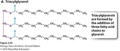
Triacylglycerols: Storage form of fat, composed of glycerol and three fatty acids.
Phospholipids: Major component of cell membranes, amphipathic with hydrophilic heads and hydrophobic tails.
Fatty Acids: Structure and Types
Fatty acids are classified based on the presence or absence of double bonds in their hydrocarbon chains.
Saturated fatty acids: No double bonds; all carbons are "full" of hydrogens. Example: butter.
Unsaturated fatty acids: One or more double bonds; causes a "kink" in the tail, increasing fluidity. Example: olive oil.
Hydrophobicity: Fatty acids are uncharged and hydrophobic, making them insoluble in water.
Example: Palmitic acid is a saturated fatty acid. 
Triacylglycerols (Triglycerides)
Triacylglycerols are formed by the addition of three fatty acid chains to a glycerol backbone. They serve as the primary energy storage molecules in animals.
Structure: Glycerol + 3 fatty acids.
Function: Energy storage.
Phospholipids and Membrane Formation
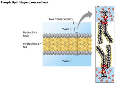
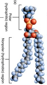
Phospholipids are amphipathic molecules, meaning they have both hydrophilic (polar) heads and hydrophobic (nonpolar) tails. This property drives the formation of the phospholipid bilayer in aqueous environments.
Hydrophilic head: Contains a phosphate group.
Hydrophobic tail: Composed of fatty acid chains.
Amphipathic nature: Essential for membrane structure.
Phospholipid Bilayer Properties
The phospholipid bilayer forms the fundamental structure of cell membranes, creating a selective barrier between the cell and its environment.
Arrangement: Hydrophilic heads face outward toward water; hydrophobic tails face inward, away from water.
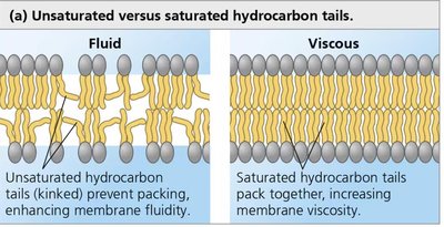
Fluidity: Unsaturated hydrocarbon tails (with kinks) prevent tight packing, increasing membrane fluidity. Saturated tails pack tightly, making the membrane more viscous.
Membrane Functions
Cell membranes serve several critical functions:
Barrier: Separates the inside of the cell from the outside environment.
Defines intracellular spaces: Allows for specialized functions within organelles.
Selective permeability: Controls what crosses the membrane.
Membrane Properties: Fluid Mosaic Model
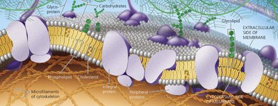
The fluid mosaic model describes membranes as dynamic structures composed of a mosaic of lipids and proteins.
Fluidity: Lipids move laterally within the bilayer; flipping is rare and enzyme-driven.
Mosaic: Proteins provide specialized functions and are distributed throughout the membrane.
Membrane Permeability and Transport
Permeability of the Phospholipid Bilayer
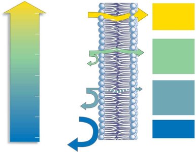
The phospholipid bilayer is selectively permeable, allowing certain molecules to cross more easily than others.
High permeability: Small, nonpolar molecules (O2, CO2, N2).
Moderate permeability: Small, uncharged polar molecules (H2O, glycerol).
Low permeability: Large, uncharged polar molecules (glucose, sucrose) and small ions (Na+, K+, Cl-).
Transport Proteins
Transport proteins facilitate the movement of molecules across the membrane, especially those that cannot diffuse freely.
Channel proteins: Provide a pathway for specific molecules to move with their concentration gradient (facilitated diffusion).
Carrier proteins: Bind to molecules and change shape to transport them across the membrane.
Active transporters: Move molecules against their concentration gradient using energy (ATP).
Membrane Proteins: Structure and Function
Types of Membrane Proteins
Membrane proteins are classified based on their association with the membrane:
Integral membrane proteins: Span the entire membrane (transmembrane proteins).
Peripheral membrane proteins: Associated with either the internal or external side of the membrane.
Amphipathic Nature of Membrane Proteins
Many membrane proteins are amphipathic, with hydrophilic regions exposed to water and hydrophobic regions embedded in the membrane.
Hydrophilic amino acid residues: Interact with aqueous environments.
Hydrophobic amino acid residues: Interact with the lipid bilayer.
Membrane Dynamics
Lateral Mobility and Fluidity
Lipids and some proteins exhibit high rates of lateral mobility within the membrane, contributing to its fluid nature.
Lateral diffusion: Movement of lipids and proteins within the plane of the membrane.
Flippases: Enzymes that facilitate the rare flipping of lipids from one leaflet to another.
Diffusion and Osmosis
Diffusion
Diffusion is the net movement of molecules from an area of high concentration to an area of low concentration due to random motion.
Driving force: Concentration gradient.
Equilibrium: Achieved when concentrations are equal on both sides.
Osmosis
Osmosis is a special case of diffusion involving the net movement of water across a semipermeable membrane.
Hypertonic solution: Higher solute concentration, lower "free" water.
Hypotonic solution: Lower solute concentration, higher "free" water.
Isotonic solution: Equal solute concentration.
Facilitated Diffusion and Active Transport
Facilitated Diffusion
Facilitated diffusion is the movement of molecules with their concentration gradient via specific membrane proteins.
Channel proteins: Allow passage of specific molecules.
Carrier proteins: Undergo conformational changes to transport molecules.
Active Transport
Active transport moves molecules against their concentration gradient, requiring energy (usually ATP).
Primary active transport: Direct use of ATP (e.g., Na+/K+ pump).
Secondary active transport: Uses energy from concentration gradients established by primary active transport.
Protein Synthesis and Membrane Integration
Protein Synthesis Locations
Proteins are synthesized in two main locations:
Free ribosomes: Synthesize proteins for the cytoplasm.
Rough endoplasmic reticulum (RER): Synthesize proteins destined for membranes or secretion.
Secretory Pathway
Proteins processed through the secretory pathway are synthesized on the RER, then transported to the Golgi complex and their final destination (plasma membrane, lysosome, etc.).
Summary Table: Lipid Types and Functions
Lipid Type | Structure | Function |
|---|---|---|
Fatty Acid | Long hydrocarbon chain with carboxyl group | Building block, energy source |
Triacylglycerol | Glycerol + 3 fatty acids | Energy storage |
Phospholipid | Glycerol + 2 fatty acids + phosphate group | Membrane structure |
Key Equations
Area of a sphere:
Volume of a sphere:
Additional info:
Membrane fluidity is crucial for cell function, affecting transport, signaling, and protein mobility.
Transport proteins are highly specific, allowing only certain molecules to pass.
Membrane proteins can be regulated, affecting transport rates and cellular responses.
