 Indietro
Membrane Structure and Function: Chapter 7 Study Notes
Guida di studio - Note intelligenti
 Appunti personalizzati basati sui tuoi materiali, ampliati con definizioni chiave, esempi e contesto.
Membrane Structure and Function
Overview: Life at the Edge
The plasma membrane is a critical boundary that separates the living cell from its environment. It exhibits selective permeability, allowing certain substances to cross more easily than others, which is essential for maintaining cellular homeostasis.
Selective permeability: Only specific molecules can pass through the membrane efficiently.
Surface area to volume ratio: Influences the efficiency of material exchange.
Concept 7.1: Cellular Membranes are Fluid Mosaics of Lipids and Proteins
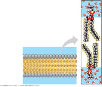
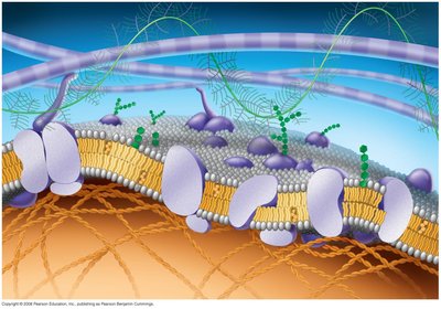
Cellular membranes are composed primarily of phospholipids and proteins, forming a fluid mosaic model. Phospholipids are amphipathic, containing both hydrophobic and hydrophilic regions, which allows them to form bilayers in aqueous environments.
Phospholipids: Most abundant lipid in the plasma membrane; amphipathic nature enables bilayer formation.
Fluid mosaic model: Membrane is a fluid structure with a mosaic of proteins embedded within.
The Fluidity of Membranes
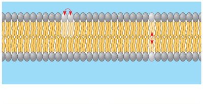
Membrane fluidity is influenced by the movement of phospholipids and proteins within the bilayer. Most lipids and some proteins drift laterally, while flip-flop movement across the membrane is rare.
Lateral movement: Occurs frequently, maintaining membrane fluidity.
Flip-flop movement: Rare, involves movement from one leaflet to another.
Factors Influencing Membrane Fluidity
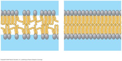
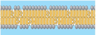
Membrane fluidity is affected by temperature, lipid composition, and cholesterol content.
Temperature: Lower temperatures cause membranes to solidify; higher temperatures increase fluidity.
Lipid composition: Unsaturated fatty acids (with kinks) increase fluidity; saturated fatty acids make membranes more viscous.
Cholesterol: At warm temperatures, cholesterol restrains phospholipid movement; at cool temperatures, it prevents tight packing, maintaining fluidity.
Membrane Proteins and Their Functions
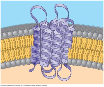
Membrane proteins are embedded in the lipid bilayer and are responsible for most of the membrane's specific functions. They are classified as integral (penetrate the hydrophobic core) or peripheral (bound to the membrane surface).
Integral proteins: Span the membrane; often called transmembrane proteins.
Peripheral proteins: Attach to the membrane surface.
Functions: Define boundaries, enzymatic activity, transport, signal transduction, cell-cell communication.
Concept 7.2: Membrane Structure Results in Selective Permeability
The plasma membrane regulates the cell's molecular traffic, allowing hydrophobic molecules to pass rapidly while polar molecules cross less easily.
Hydrophobic molecules: Pass through the lipid bilayer quickly (e.g., hydrocarbons).
Polar molecules: Cross the membrane slowly (e.g., sugars).
Concept 7.3: Passive Transport Across Membranes
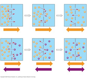
Passive transport involves the diffusion of substances across a membrane without energy investment. Molecules move from areas of high concentration to low concentration until dynamic equilibrium is reached.
Diffusion: Movement of molecules from high to low concentration.
Dynamic equilibrium: Equal movement in both directions.
Osmosis and Water Balance
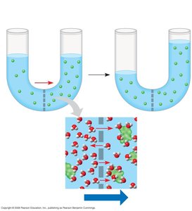
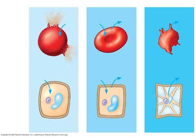
Osmosis is the diffusion of water across a selectively permeable membrane. Water moves from regions of lower solute concentration to higher solute concentration.
Isotonic solution: Equal solute concentration inside and outside the cell; no net water movement.
Hypertonic solution: Higher solute concentration outside; cell loses water.
Hypotonic solution: Lower solute concentration outside; cell gains water.
Transport Proteins
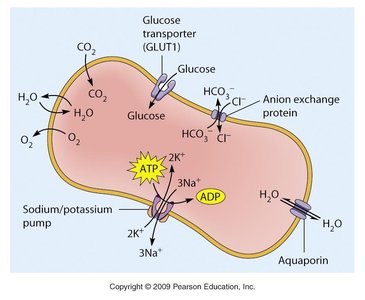
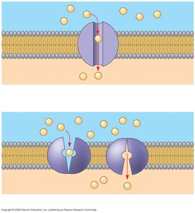
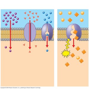
Transport proteins facilitate the movement of hydrophilic substances across the membrane. Channel proteins provide corridors, while carrier proteins bind and shuttle molecules across.
Channel proteins: Form hydrophilic channels for specific molecules or ions.
Carrier proteins: Bind to molecules and change shape to transport them.
Specificity: Each transport protein is specific for the substance it moves.
Concept 7.4: Active Transport
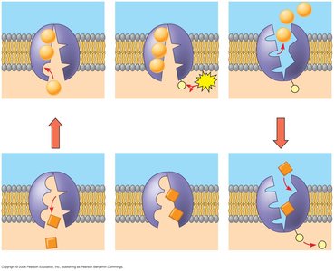
Active transport moves substances against their concentration gradient and requires energy, usually in the form of ATP. This process allows cells to maintain concentration gradients that differ from their surroundings.
Active transport: Requires energy input; moves substances from low to high concentration.
Example: Sodium-potassium pump maintains Na+ and K+ gradients across the membrane.
Summary of Transport Mechanisms
Type | Energy Required | Direction |
|---|---|---|
Diffusion | No | High to Low |
Facilitated Diffusion | No | High to Low |
Active Transport | Yes (ATP) | Low to High |
Membrane Carbohydrates and Cell-Cell Communication
Cells recognize each other by binding to surface molecules, often carbohydrates, on the plasma membrane. These carbohydrates may be covalently bonded to lipids (glycolipids) or proteins (glycoproteins), and their composition varies among species and cell types.
Glycolipids: Carbohydrates attached to lipids.
Glycoproteins: Carbohydrates attached to proteins.
Concept 7.5: Bulk Transport Across the Plasma Membrane
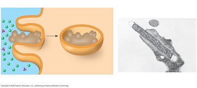
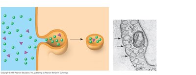
Bulk transport involves the movement of large molecules across the membrane via vesicles. This process requires energy and includes exocytosis and endocytosis.
Exocytosis: Transport vesicles fuse with the membrane to release contents outside the cell.
Endocytosis: Cell takes in macromolecules by forming vesicles from the plasma membrane.
Types of endocytosis: Phagocytosis (cellular eating), pinocytosis (cellular drinking), receptor-mediated endocytosis.
Cystic Fibrosis: Membrane Transport Disease Example
CFTR Protein and Cystic Fibrosis
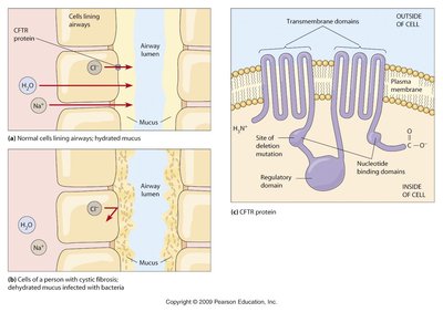
Cystic fibrosis is caused by mutations in the CFTR protein, a membrane transport protein. In normal cells, CFTR regulates the movement of chloride ions and water, maintaining hydrated mucus in airways. In cystic fibrosis, defective CFTR leads to dehydrated mucus and increased susceptibility to bacterial infections.
CFTR protein: Transmembrane protein involved in chloride ion transport.
Normal function: Maintains hydration of airway mucus.
Defective function: Results in thick, dehydrated mucus and infection risk.
Hemolysins and Membrane Disruption
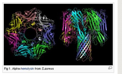
Hemolysins are proteins produced by bacteria such as Staphylococcus aureus that disrupt cell membranes, leading to cell lysis. These proteins form pores in the membrane, compromising its integrity.
Hemolysins: Bacterial proteins that lyse host cells by forming membrane pores.
Example: Alpha-hemolysin from S. aureus.
Chapter 7 Study Guide: Key Concepts
Membrane fluidity is influenced by temperature, cholesterol, and membrane composition (saturated vs. unsaturated fatty acids).
Distinguish between peripheral and integral membrane proteins; osmosis, facilitated diffusion, passive and active transport; hypertonic, hypotonic, and isotonic solutions.
Understand the five functions of biological membranes: boundary definition, enzymatic activity, transport, signal transduction, and cell-cell communication.
Explain how large molecules are transported across the cell membrane via exocytosis and endocytosis.
Additional info: Academic context was added to clarify mechanisms, definitions, and examples for each topic.
