Indietro
Membrane Structure and Function: Study Guide for General Biology
Guida di studio - Note intelligenti
 Appunti personalizzati basati sui tuoi materiali, ampliati con definizioni chiave, esempi e contesto.
Membrane Structure and Function
Overview of Cellular Membranes
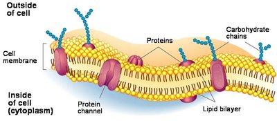
Cellular membranes are essential for maintaining homeostasis and regulating the internal environment of the cell. They are composed primarily of lipids and proteins, with carbohydrates playing important roles in cell recognition and signaling.
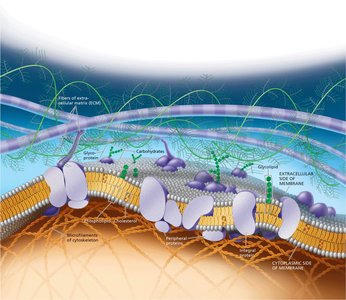
Fluid Mosaic Model: The cell membrane is a dynamic structure where protein molecules drift laterally in a fluid bilayer of phospholipids.
Main Components: Lipids (especially phospholipids), proteins, and carbohydrates.
Phospholipids: Most abundant lipid; their amphipathic nature allows them to form bilayers.
Phospholipids and Amphipathic Nature
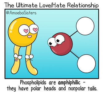
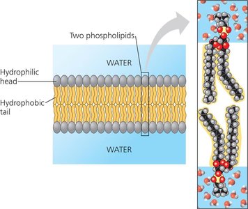
Phospholipids are amphipathic molecules, meaning they have both hydrophilic (water-loving) heads and hydrophobic (water-fearing) tails. This property is crucial for the formation of the bilayer structure of membranes.
Hydrophilic Head: Attracts water, faces outward toward aqueous environments.
Hydrophobic Tail: Repels water, faces inward, away from water.
Membrane Proteins
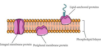
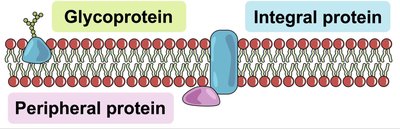
Proteins embedded in the membrane are also often amphipathic and are not randomly distributed. They play key roles in transport, signaling, and structural support.
Integral Proteins: Penetrate the hydrophobic interior; most are transmembrane proteins.
Peripheral Proteins: Loosely bound to the membrane surface or to integral proteins.
Lipid-Anchored Proteins: Covalently attached to lipids within the membrane.
Fluidity of Membranes
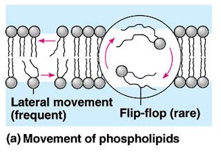
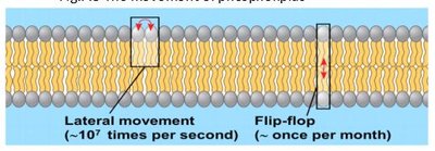
The fluidity of the membrane is maintained by hydrophobic interactions, which are weaker than covalent bonds. Lipids and some proteins can move laterally within the membrane, while flip-flop movement across the bilayer is rare.
Lateral Movement: Frequent and rapid for phospholipids.
Flip-Flop Movement: Rare, occurs once per month for a lipid molecule.
Factors Affecting Membrane Fluidity
Membrane fluidity is influenced by temperature, cholesterol, and the saturation of fatty acid tails.
Temperature: Lower temperatures can cause membranes to solidify.
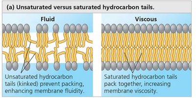
Unsaturated Fatty Acids: Increase fluidity due to kinks in tails preventing tight packing.
Saturated Fatty Acids: Decrease fluidity by allowing tight packing.
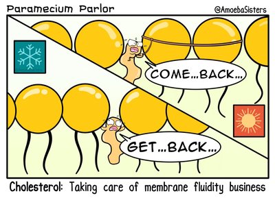
Cholesterol: Acts as a fluidity buffer, maintaining membrane fluidity across temperature changes.
Evolutionary Adaptations in Membrane Lipid Composition
Organisms adapt their membrane lipid composition to environmental conditions to maintain proper fluidity.
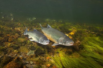
Cold Environments: More unsaturated hydrocarbon tails (e.g., fish in cold water).
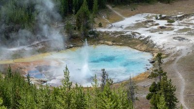
Hot Environments: More saturated fatty acids (e.g., bacteria in hot springs).
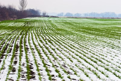
Plants: Increase unsaturated phospholipids in cold seasons to prevent membrane solidification.
Membrane Proteins and Their Functions
Membrane proteins are responsible for most of the functions of the membrane, including transport, enzymatic activity, signal transduction, cell-cell recognition, intercellular joining, and attachment to the cytoskeleton and extracellular matrix.
Integral Proteins: Span the membrane and are involved in transport and signaling.
Peripheral Proteins: Provide structural support and participate in cell signaling.
The Role of Membrane Carbohydrates in Cell-Cell Recognition
Membrane carbohydrates are crucial for cell-cell recognition, allowing cells to distinguish neighboring cells and facilitating immune responses.
Glycolipids: Carbohydrates covalently bonded to lipids.
Glycoproteins: Carbohydrates covalently bonded to proteins; most common.
Blood Types: Human blood types (A, B, AB, O) are determined by variations in glycoproteins on red blood cells.
Membrane Structure and Selective Permeability
Biological membranes exhibit selective permeability, allowing the cell to regulate the passage of substances.
Cross Easily: Small nonpolar molecules (hydrocarbons, CO2, O2).
Not as Easily: Polar molecules (water, glucose).
Unlikely to Cross: Ions (charged molecules).
Transport Proteins
Transport proteins facilitate the movement of hydrophilic substances across the membrane.
Channel Proteins: Provide hydrophilic channels for molecules or ions.
Carrier Proteins: Change shape to shuttle substances across the membrane.
Specificity: Each transport protein is specific for the substance it moves.
Passive Transport
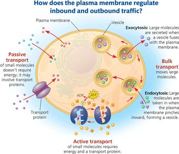
Passive transport involves the diffusion of substances across the membrane without energy expenditure.
Diffusion: Movement from high to low concentration.
Concentration Gradient: Each substance diffuses down its own gradient.
Osmosis: Diffusion of water across a selectively permeable membrane.
Water Balance of Cells Without a Cell Wall
Animal cells must regulate water balance to prevent excessive uptake or loss of water.
Isotonic Solution: No net movement of water; cell volume is stable.
Hypertonic Solution: Cell loses water, shrivels, and may die.
Hypotonic Solution: Cell gains water, swells, and may burst.
Osmoregulation: Control of solute concentrations and water balance.
Water Balance of Cells With a Cell Wall
Plant, prokaryote, and fungal cells have cell walls that help maintain water balance.
Turgid: Swollen state; healthy for most plants.
Flaccid: Limp state; occurs in isotonic solutions.
Plasmolysis: Cytoplasm shrivels and membrane pulls away from cell wall in hypertonic solutions.
Facilitated Diffusion
Facilitated diffusion is passive transport aided by proteins, allowing molecules or ions to move down their concentration gradient efficiently.
Ion Channels: Many function as gated channels, opening or closing in response to stimuli.
Active Transport
Active transport moves substances against their concentration gradients, requiring energy input, usually from ATP.
Carrier Proteins: Mediate active transport.
Example: Sodium-potassium pump maintains high K+ and low Na+ inside animal cells.
ATP Hydrolysis: Provides energy by transferring a phosphate group to the transport protein, inducing a shape change.
Membrane Potential and Ion Pumps
All cells have voltages across their plasma membranes, known as membrane potential, which is crucial for transport and signaling.
Electrochemical Gradient: Combination of chemical and electrical forces driving ion diffusion.
Electrogenic Pumps: Generate voltage across membranes (e.g., sodium-potassium pump in animals, proton pump in plants, fungi, bacteria).
Cotransport
Cotransport involves the coupling of the downhill diffusion of one substance to the uphill transport of another, using gradients established by active transport.
Example: Plant cells use proton gradients to drive active transport of nutrients.
Bulk Transport: Exocytosis and Endocytosis
Large molecules enter and leave the cell in bulk via vesicles, through processes called exocytosis and endocytosis.
Exocytosis: Secretion of biological molecules by fusion of vesicles with the plasma membrane.
Endocytosis: Uptake of molecules by formation of vesicles from the plasma membrane.
Phagocytosis: "Cellular eating"; cell engulfs large particles.
Pinocytosis: "Cellular drinking"; cell ingests extracellular fluid and solutes.
Receptor-Mediated Endocytosis: Specific molecules are taken in via vesicles with receptor sites.
Key Terms and Definitions
Amphipathic: Molecule with both hydrophilic and hydrophobic regions.
Selective Permeability: Ability of membrane to regulate passage of substances.
Osmosis: Diffusion of water across a membrane.
Tonicity: Ability of a solution to cause a cell to gain or lose water.
Membrane Potential: Voltage across a membrane.
Electrochemical Gradient: Combined effect of concentration and electrical gradients on ion movement.
Important Equations
Diffusion: $ J = -D \frac{dC}{dx} $ Where J is the flux, D is the diffusion coefficient, and $ \frac{dC}{dx} $ is the concentration gradient.
Membrane Potential: $ V_m = V_{inside} - V_{outside} $ Where $ V_m $ is membrane potential.
Summary Table: Types of Membrane Transport
Type | Energy Required? | Direction | Example |
|---|---|---|---|
Passive Transport | No | Down gradient | O2 diffusion |
Facilitated Diffusion | No | Down gradient | Glucose via carrier protein |
Active Transport | Yes (ATP) | Against gradient | Na+/K+ pump |
Bulk Transport | Yes (vesicles) | In/out of cell | Exocytosis, endocytosis |