Indietro
Membrane Structure and Function: Study Notes for General Biology
Guida di studio - Note intelligenti
 Appunti personalizzati basati sui tuoi materiali, ampliati con definizioni chiave, esempi e contesto.
Membrane Structure and Function
Overview of the Cell Membrane
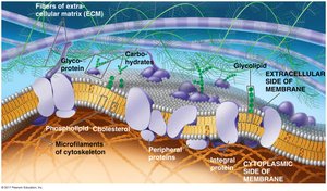
The cell membrane, also known as the plasma membrane, is a dynamic structure that separates the interior of the cell from its external environment. It is primarily composed of a lipid bilayer with embedded proteins, carbohydrates, and cholesterol, each contributing to its function and fluidity.
Lipid Bilayer: The fundamental structure of the membrane, consisting of two layers of phospholipids with hydrophobic tails facing inward and hydrophilic heads facing outward.
Proteins: Integral and peripheral proteins are embedded or attached to the membrane, performing functions such as transport, signaling, and structural support.
Carbohydrates: Often attached to proteins (glycoproteins) or lipids (glycolipids), these molecules are involved in cell recognition and communication.
Cholesterol: Interspersed within the bilayer, cholesterol modulates membrane fluidity and stability.
Functions of Membrane Proteins
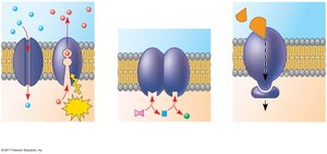
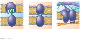
Membrane proteins are essential for the diverse functions of the cell membrane. They can be classified based on their roles:
Transport: Proteins facilitate the movement of substances across the membrane, including channel and carrier proteins.
Enzymatic Activity: Some membrane proteins act as enzymes, catalyzing reactions at the membrane surface.
Signal Transduction: Receptor proteins receive signaling molecules and initiate cellular responses.
Cell-Cell Recognition: Glycoproteins serve as identification tags for cell recognition.
Intercellular Joining: Proteins form junctions between cells for communication and adhesion.
Attachment to Cytoskeleton and ECM: Proteins anchor the membrane to internal and external structural components.
Membrane Fluidity and the Fluid Mosaic Model
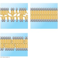
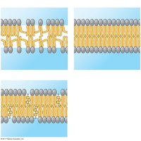
The cell membrane is described by the fluid mosaic model, which emphasizes its dynamic nature and the lateral movement of components. Membrane fluidity is influenced by lipid composition and temperature.
Unsaturated vs. Saturated Fatty Acids: Unsaturated hydrocarbon tails prevent tight packing, increasing fluidity, while saturated tails pack closely, making the membrane more viscous.
Cholesterol: Acts as a fluidity buffer, reducing fluidity at high temperatures and preventing solidification at low temperatures.
Movement of Materials Across the Membrane
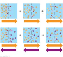
The cell membrane is selectively permeable, allowing certain substances to pass while restricting others. Transport mechanisms include passive and active processes.
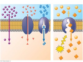
Diffusion: The movement of molecules from an area of higher concentration to lower concentration without energy input.
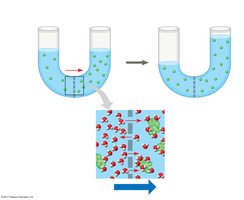
Osmosis: The diffusion of water across a selectively permeable membrane.
Facilitated Diffusion: Passive transport aided by proteins, such as channel and carrier proteins.
Active Transport: Movement of substances against their concentration gradient, requiring energy (ATP).
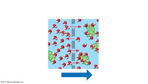
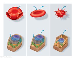
Tonicity and Water Balance in Cells
Tonicity refers to the ability of a solution to cause a cell to gain or lose water, depending on the concentration of solutes that cannot cross the membrane.
Hypotonic Solution: Lower solute concentration outside the cell; water enters, causing the cell to swell.
Isotonic Solution: Equal solute concentration; no net water movement.
Hypertonic Solution: Higher solute concentration outside; water leaves, causing the cell to shrink.
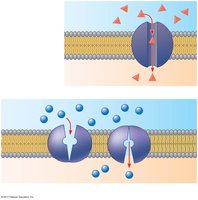
Facilitated Diffusion: Channel and Carrier Proteins
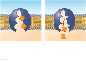
Facilitated diffusion is a type of passive transport where proteins assist in moving substances across the membrane. Channel proteins create pores for specific molecules, while carrier proteins undergo conformational changes to transport substances.
Aquaporins: Channel proteins specifically for water.
Ion Channels: Allow ions to pass through the membrane.
Glucose Transporters: Carrier proteins for glucose.
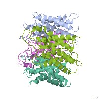
Active Transport: Sodium-Potassium Pump
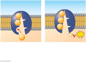
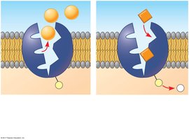
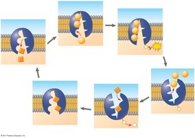
Active transport uses energy to move substances against their concentration gradients. The sodium-potassium pump is a classic example, maintaining cellular ion balance by pumping Na+ out and K+ in.
Mechanism: Involves ATP hydrolysis, phosphorylation, and conformational changes in the pump protein.
Importance: Maintains membrane potential and cellular homeostasis.
Passive vs. Active Transport
Transport proteins can facilitate both passive and active transport. Passive transport does not require energy, while active transport does.
Passive Transport: Includes diffusion and facilitated diffusion.
Active Transport: Requires ATP and moves substances against their gradient.
Electrochemical Gradient and Membrane Potential
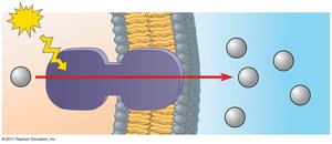
The membrane potential is the voltage across the membrane, created by differences in ion concentrations. The electrochemical gradient drives ion diffusion based on both chemical and electrical forces.
Proton Pumps: Actively transport H+ ions, contributing to membrane potential.
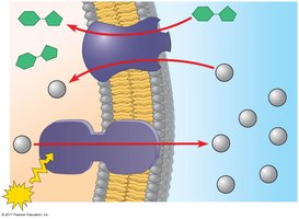
Cotransport: Active transport of one solute indirectly drives the transport of another.
Bulk Transport: Endocytosis and Exocytosis
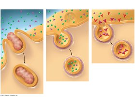
Bulk transport moves large molecules or particles across the membrane via vesicles. Endocytosis brings substances into the cell, while exocytosis expels them.
Phagocytosis: Cell engulfs large particles.
Pinocytosis: Cell takes in fluid and dissolved solutes.
Receptor-Mediated Endocytosis: Specific molecules are taken in after binding to receptors.
Membrane Trafficking in the Cell
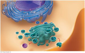
Membrane trafficking involves the movement of vesicles between cellular compartments, such as the Golgi apparatus, endoplasmic reticulum, and plasma membrane. This process is essential for secretion, endocytosis, and membrane renewal.