 Indietro
Membrane Structure and Function: The Fluid Mosaic Model and Membrane Transport
Guida di studio - Note intelligenti
 Appunti personalizzati basati sui tuoi materiali, ampliati con definizioni chiave, esempi e contesto.
Plasma Membrane: Structure and Function
Overview of the Plasma Membrane
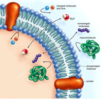
The plasma membrane is the boundary that separates the living cell from its external environment. It plays a critical role in regulating the exchange of substances, maintaining homeostasis, and facilitating communication between cells. The plasma membrane exhibits selective permeability, allowing some substances to cross more easily than others.
Selective permeability: Only certain molecules can pass through freely; others require assistance.
Regulation: Controls the transport of small and macromolecules, and the flow of information between cells.
Cell adhesion: Facilitates tissue formation and cellular recognition.
Components of the Plasma Membrane
The plasma membrane is composed of several key components, each contributing to its structure and function:
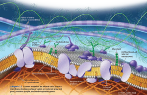
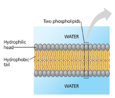
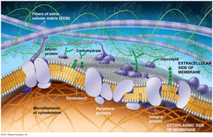
Phospholipid bilayer: Forms the fundamental structure, with hydrophilic heads facing outward and hydrophobic tails inward.
Membrane proteins: Integral and peripheral proteins serve roles in transport, signaling, and structural support.
Carbohydrates: Attached to proteins (glycoproteins) or lipids (glycolipids), important for cell recognition.
Cholesterol: Modulates membrane fluidity and stability.
Fluid Mosaic Model
Structure and Properties
The fluid mosaic model describes the plasma membrane as a dynamic structure where lipids and proteins move laterally within the layer, creating a flexible yet organized arrangement. The membrane's fluidity is essential for its function, including transport and cell signaling.
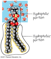
Amphipathic molecules: Phospholipids have both hydrophilic (water-loving) heads and hydrophobic (water-fearing) tails.
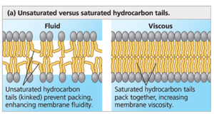
Fluidity: Determined by the degree of unsaturation of fatty acids and cholesterol content.
Mosaic: Proteins are interspersed throughout the lipid bilayer, creating a mosaic pattern.
Factors Affecting Membrane Fluidity
Fatty acid tail length: Shorter tails increase fluidity; longer tails decrease fluidity.
Degree of saturation: Unsaturated fatty acids (with double bonds) increase fluidity; saturated fatty acids decrease fluidity and increase viscosity.
Cholesterol: Acts as a "fluidity buffer," preventing the membrane from becoming too rigid or too fluid.
Temperature: High temperatures increase fluidity; low temperatures decrease fluidity.
Membrane Proteins and Their Functions
Types of Membrane Proteins
Integral proteins: Penetrate the hydrophobic core of the lipid bilayer; most are transmembrane proteins that span the membrane. They often function as transport proteins, channels, or carriers.
Peripheral proteins: Loosely bound to the membrane surface or to integral proteins; involved in support, signaling, and maintaining cell shape.
Functions of Membrane Proteins
Transport: Channel and carrier proteins facilitate the movement of substances across the membrane.
Enzymatic activity: Some proteins act as enzymes, catalyzing metabolic reactions.
Signal transduction: Receptor proteins bind signaling molecules and initiate cellular responses.
Cell-cell recognition: Glycoproteins serve as identification tags for cellular recognition.
Intercellular joining: Proteins form junctions between adjacent cells.
Attachment to cytoskeleton and ECM: Maintains cell shape and stabilizes protein positions.
Membrane Carbohydrates
Role in Cell Recognition
Membrane carbohydrates are short, branched chains of sugars attached to lipids (glycolipids) or proteins (glycoproteins). They play a crucial role in cell-cell recognition, tissue formation, and immune response.
Glycoproteins and glycolipids: Serve as markers for cellular identification and sorting during development.
Membrane Transport Mechanisms
Passive Transport
Passive transport involves the movement of substances across the membrane without energy input, down their concentration gradient.
Simple diffusion: Movement of small, nonpolar molecules (e.g., O2, CO2) directly through the lipid bilayer.
Facilitated diffusion: Movement of polar or charged molecules via specific transport proteins (channels or carriers).
Osmosis: Diffusion of water through a selectively permeable membrane, often via aquaporins.
Fick's Law of Diffusion:
Factors affecting diffusion rate: Molecule size, temperature, surface area, and concentration gradient.
Selective Permeability
Nonpolar molecules: Diffuse easily through the membrane.
Polar molecules and ions: Require transport proteins to cross the membrane.
Water: Moves via aquaporins (channel proteins).
Facilitated Diffusion
Channel proteins: Provide hydrophilic pathways for specific molecules or ions (e.g., aquaporins for water, ion channels for Na+, K+).
Carrier proteins: Bind and transport specific molecules by changing shape.
Osmosis and Tonicity
Osmosis: Movement of water from high to low concentration through a selectively permeable membrane.
Tonicity: The ability of a solution to cause a cell to gain or lose water.
Hypertonic solution: Higher solute concentration; cell loses water.
Hypotonic solution: Lower solute concentration; cell gains water.
Isotonic solution: Equal solute concentration; no net water movement.
Water Balance in Cells
Animal cells: Sensitive to changes in tonicity; may burst or shrink.
Plant cells: Cell wall provides support; turgor pressure maintains rigidity in hypotonic environments.
Active Transport
Mechanisms and Examples
Active transport requires energy (usually ATP) to move substances against their concentration gradient. This process is essential for maintaining cellular homeostasis.
Sodium-potassium pump: Moves Na+ out and K+ into the cell, crucial for nerve impulse transmission.
Proton pump: Transports H+ ions across membranes, important in cellular respiration and photosynthesis.
Cotransport (secondary active transport): One substance moves down its gradient, providing energy to transport another substance against its gradient (e.g., antiporters).
Bulk Transport: Endocytosis and Exocytosis
Endocytosis
Phagocytosis: "Cellular eating"; intake of solid particles.
Pinocytosis: "Cellular drinking"; intake of extracellular fluid.
Receptor-mediated endocytosis: Specific molecules are taken in after binding to receptors.
Exocytosis
Discharge of materials from vesicles at the cell surface (e.g., secretion of mucus by goblet cells).
Additional info: The notes above integrate and expand upon the provided material, ensuring a comprehensive, self-contained summary suitable for college-level General Biology students. Images were included only when directly relevant to the adjacent explanation.
