 Indietro
Membrane Structure and Function: The Working Cell
Guida di studio - Note intelligenti
 Appunti personalizzati basati sui tuoi materiali, ampliati con definizioni chiave, esempi e contesto.
Membrane Structure and Function
Fluid Mosaic Model of Membrane Structure
The cell membrane is a dynamic structure described by the fluid mosaic model. This model explains how membranes are composed of a phospholipid bilayer with embedded proteins, cholesterol, and carbohydrates, allowing for flexibility and diverse functions.
Phospholipids form the basic structure, creating a bilayer with hydrophilic heads facing outward and hydrophobic tails inward.
Proteins are interspersed throughout the bilayer, serving various roles such as transport, signaling, and structural support.
Cholesterol is embedded within the bilayer, contributing to membrane fluidity and stability.
Carbohydrates are attached to proteins (glycoproteins) and lipids (glycolipids), functioning in cell recognition.
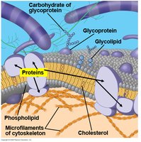
Additional info: The fluid mosaic model allows for lateral movement of components, which is essential for membrane function and cell signaling.
Membrane Fluidity
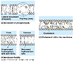
Membrane fluidity is determined by the composition of phospholipids and the presence of cholesterol. Unsaturated fatty acids in phospholipids create kinks, preventing tight packing and increasing fluidity. Cholesterol acts as a buffer, maintaining fluidity at various temperatures.
Unsaturated fatty acids increase fluidity due to kinks in their hydrocarbon tails.
Saturated fatty acids decrease fluidity by allowing tight packing.
Cholesterol stabilizes the membrane, preventing it from becoming too fluid or too rigid.
Phospholipid Bilayer Structure
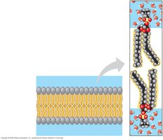
The phospholipid bilayer is arranged with hydrophilic (polar) heads facing the aqueous environment and hydrophobic (nonpolar) tails facing inward, creating a selective barrier.
Hydrophilic heads interact with water inside and outside the cell.
Hydrophobic tails avoid water, forming the interior of the membrane.
This arrangement is crucial for selective permeability, allowing only certain molecules to pass.
Membrane Proteins and Their Functions
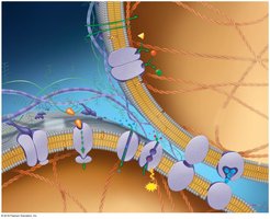
Membrane proteins are essential for the diverse functions of the cell membrane. They are classified based on their roles:
Transport proteins: Facilitate the movement of substances across the membrane.
Enzymatic proteins: Catalyze chemical reactions at the membrane surface.
Receptor proteins: Receive and transmit signals from the environment.
Glycoproteins: Serve as identification tags for cell recognition.
Junction proteins: Connect adjacent cells, forming tissues.
Attachment proteins: Anchor the membrane to the cytoskeleton and extracellular matrix.
Membrane Permeability and Transport
Selective Permeability
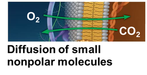
The cell membrane exhibits selective permeability, allowing some substances to cross more easily than others. Small, nonpolar molecules (e.g., O2, CO2) can diffuse directly through the bilayer, while polar or charged molecules require transport proteins.
Direct diffusion: Small, nonpolar molecules pass freely.
Transport proteins: Facilitate movement of ions, sugars, and other polar molecules.
Passive Transport
Passive transport involves the movement of substances across the membrane without energy expenditure. Types include:
Simple diffusion: Movement from high to low concentration.
Osmosis: Diffusion of water across a selectively permeable membrane.
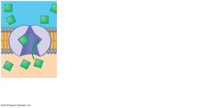
Facilitated diffusion: Movement of substances via transport proteins, still from high to low concentration.
Example: Oxygen and carbon dioxide diffuse directly through the membrane, while glucose requires a transport protein.
Osmosis and Water Balance
Osmosis is the diffusion of water across a membrane in response to solute concentration differences. Water moves from areas of low solute concentration to high solute concentration until equilibrium is reached.
Isotonic solution: Equal solute concentration; no net water movement.
Hypotonic solution: Lower solute concentration; water enters the cell.
Hypertonic solution: Higher solute concentration; water leaves the cell.
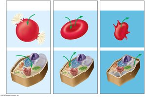
Additional info: Plant cells become turgid in hypotonic solutions due to cell wall pressure, while animal cells may lyse.
Facilitated Diffusion and Aquaporins
Facilitated diffusion allows specific molecules to cross the membrane with the help of transport proteins. Aquaporins are specialized channels for rapid water movement.
Transport proteins are specific for certain molecules (e.g., glucose, ions).
Aquaporins enable efficient water transport, crucial for osmoregulation.

Active Transport
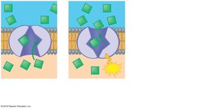
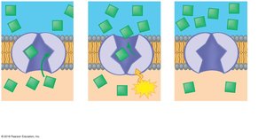
Active transport requires energy (ATP) to move substances against their concentration gradient. This process involves transport proteins that change shape upon phosphorylation.
Example: Sodium-potassium pump moves Na+ out and K+ into the cell.
Equation: (energy release for transport)
Bulk Transport: Exocytosis and Endocytosis
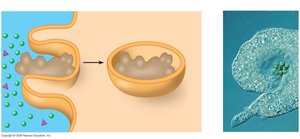
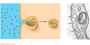
Cells use exocytosis and endocytosis to transport large molecules. Exocytosis exports materials by vesicle fusion with the membrane, while endocytosis imports substances by engulfing them.
Phagocytosis: Cell engulfs particles, forming a vacuole.
Pinocytosis: Cell takes in fluid via small vesicles.
Receptor-mediated endocytosis: Specific molecules are taken in after binding to membrane receptors.
Summary Table: Membrane Transport Mechanisms
Transport Type | Energy Required | Direction | Example |
|---|---|---|---|
Simple Diffusion | No | High to Low | O2, CO2 |
Facilitated Diffusion | No | High to Low | Glucose, ions |
Osmosis | No | High to Low (water) | Water movement |
Active Transport | Yes (ATP) | Low to High | Na+/K+ pump |
Exocytosis | Yes | Out of cell | Protein secretion |
Endocytosis | Yes | Into cell | Phagocytosis, pinocytosis |
