 Indietro
Membrane Transport and Cell Signaling: Study Notes for General Biology
Guida di studio - Note intelligenti
 Appunti personalizzati basati sui tuoi materiali, ampliati con definizioni chiave, esempi e contesto.
Membrane Structure and Function
Fluid Mosaic Model of Cellular Membranes
The plasma membrane is a dynamic structure composed of lipids, proteins, and carbohydrates, forming a fluid mosaic that regulates cellular interactions and transport.
Phospholipids are amphipathic, containing both hydrophilic heads and hydrophobic tails, forming a bilayer that acts as a stable boundary between aqueous compartments.
Membrane proteins include integral proteins (spanning the bilayer) and peripheral proteins (attached to the membrane surface).
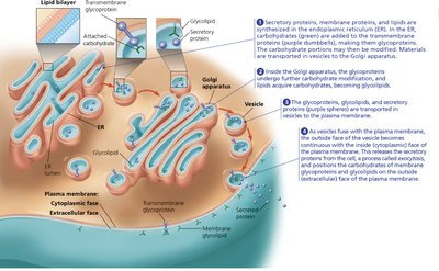
Membrane carbohydrates are involved in cell-cell recognition, often attached to proteins (glycoproteins) or lipids (glycolipids).
The fluid mosaic model describes the membrane as a mosaic of proteins bobbing in a fluid phospholipid bilayer.
Example: The asymmetric arrangement of proteins, lipids, and carbohydrates is established during membrane synthesis in the ER and Golgi apparatus.
Membrane Transport
Selective Permeability
Cell membranes allow some substances to cross more easily than others, maintaining internal homeostasis.
Hydrophobic molecules (e.g., hydrocarbons) can dissolve in the lipid bilayer and cross easily.
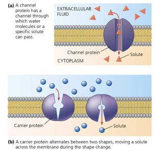
Hydrophilic molecules (e.g., ions, polar molecules) require transport proteins to cross the membrane.
Transport proteins include channel proteins (form pores) and carrier proteins (change shape to move substances).
Passive and Active Transport
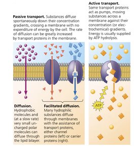
Transport across membranes can be passive (no energy required) or active (requires energy).
Passive transport includes diffusion and facilitated diffusion, moving substances down their concentration gradient.
Active transport uses energy (usually ATP) to move substances against their concentration gradient, often via carrier proteins.
Facilitated diffusion is aided by channel or carrier proteins, allowing hydrophilic substances to cross the membrane.
Cotransport and Proton Pumps
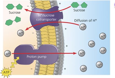
Cotransport involves the coupling of the movement of one substance down its gradient with the movement of another substance against its gradient.
Proton pumps use ATP to create a gradient of H+ ions across the membrane.
Cotransporters use the energy stored in the H+ gradient to transport other molecules (e.g., sucrose) into the cell.
Bulk Transport: Exocytosis and Endocytosis
Exocytosis
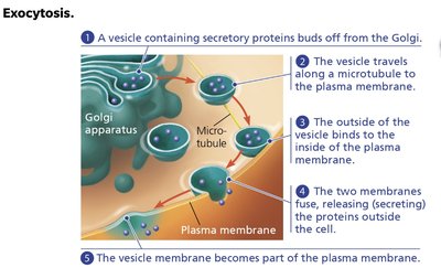
Exocytosis is the process by which cells secrete large molecules by the fusion of vesicles with the plasma membrane.
Secretory vesicles bud from the Golgi apparatus and travel along microtubules to the plasma membrane.
Fusion of the vesicle and plasma membrane releases the contents outside the cell.
The vesicle membrane becomes part of the plasma membrane.
Endocytosis
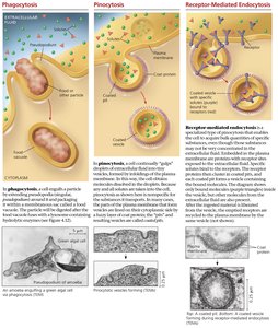
Endocytosis is the process by which cells take in molecules and particulate matter by forming new vesicles from the plasma membrane.
Phagocytosis: Cell engulfs large particles or organisms.
Pinocytosis: Cell ingests extracellular fluid and dissolved solutes.
Receptor-mediated endocytosis: Cell takes in specific molecules via receptor proteins.
Cell Signaling
Local and Long-Distance Signaling
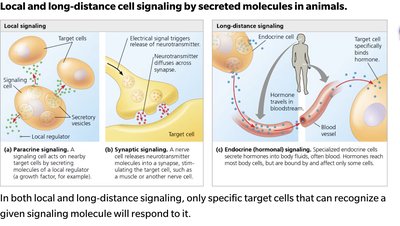
Cells communicate via signaling molecules that can act locally or over long distances.
Local signaling includes paracrine and synaptic signaling, affecting nearby cells.
Long-distance signaling involves hormones traveling through the bloodstream to target cells.
Stages of Cell Signaling
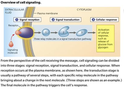
Cell signaling involves three main stages: reception, transduction, and response.
Reception: Detection of a signaling molecule by a receptor protein.
Transduction: Conversion of the signal to a form that can trigger a cellular response, often involving a signal transduction pathway.
Response: Activation of a specific cellular activity.
Receptors and Signal Transduction
Receptors can be located on the cell surface or inside the cell, and their activation initiates signal transduction pathways.
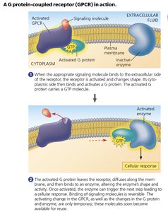
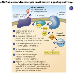
G protein-coupled receptors (GPCRs) activate G proteins, which then trigger downstream signaling events.
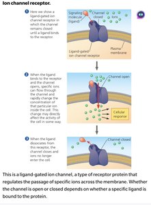
Ligand-gated ion channels open or close in response to ligand binding, allowing ions to cross the membrane.
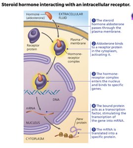
Intracellular receptors bind hydrophobic signaling molecules (e.g., steroid hormones) and often regulate gene expression.
Signal Transduction Pathways
Signal transduction often involves cascades of molecular interactions, amplifying and regulating the cellular response.
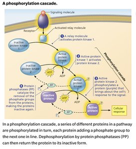
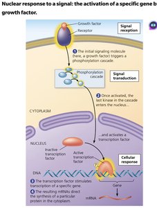
Phosphorylation cascades involve protein kinases transferring phosphate groups to other proteins, activating them.
Protein phosphatases remove phosphate groups, deactivating proteins and allowing regulation of the pathway.
Second messengers (e.g., cAMP) are small molecules that relay signals inside the cell.
Cellular Responses to Signals
Cellular responses can include changes in gene expression or alterations in cytoplasmic activities.
Signaling pathways can activate transcription factors, leading to the synthesis of specific proteins.
Other responses may involve changes in enzyme activity or opening of ion channels.
Additional info: These notes cover the essential concepts of membrane structure, transport, and cell signaling, as outlined in General Biology chapters 5 and 6. All included images directly reinforce the explanations and are relevant to the adjacent content.
