 Indietro
Microscopy and Cell Fractionation: Tools for Studying Cells
Guida di studio - Note intelligenti
 Appunti personalizzati basati sui tuoi materiali, ampliati con definizioni chiave, esempi e contesto.
Microscopy and Cell Fractionation
Introduction to Cell Study
Biologists use advanced tools and techniques to investigate cells, which are typically too small to be seen with the naked eye. The study of cells involves both microscopy and biochemical methods, allowing researchers to explore cell structure and function in detail.
Microscopy: Types and Principles
Microscopy is fundamental to cell biology, enabling visualization of cells and their components. The invention and refinement of microscopes since the 16th century have revolutionized biological research.
Light Microscopes (LM): Use visible light passed through specimens and glass lenses to magnify images. Suitable for observing living cells and tissues.
Electron Microscopes (EM): Use beams of electrons instead of light, providing much higher resolution. Two main types are:
Scanning Electron Microscope (SEM): Visualizes surface topography in three dimensions.
Transmission Electron Microscope (TEM): Reveals internal cell structures by passing electrons through thin sections.
Super-Resolution Microscopy: Recent advances allow visualization of subcellular structures beyond the traditional resolution limits.
Key Parameters:
Magnification: Ratio of image size to actual size.
Resolution: Ability to distinguish two close points as separate; inversely related to wavelength ().
Contrast: Difference in brightness between parts of the specimen.
Example: Light microscopes are ideal for studying living cells, while electron microscopes are used for detailed analysis of cell organelles.
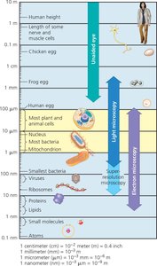
Size Range of Cells and Metric System
Cells and their components vary greatly in size, from visible objects like eggs to microscopic structures such as organelles and molecules. Understanding these sizes requires familiarity with the metric system and its prefixes.
Most plant and animal cells: 10–100 μm in diameter
Bacteria: 1–10 μm
Organelles (e.g., mitochondria): 1–10 μm
Viruses: 50–100 nm
Proteins and lipids: 1–10 nm
Metric Prefixes: Used to express measurements at different scales.
Prefix | Symbol | Exponent | Multiplier (factor) |
|---|---|---|---|
centi- | c | 10-2 | 1/100 |
milli- | m | 10-3 | 1/1000 |
micro- | μ | 10-6 | 1/1,000,000 |
nano- | n | 10-9 | 1/1,000,000,000 |
pico- | p | 10-12 | 1/1,000,000,000,000 |
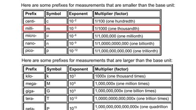
Example: A typical bacterial cell is about 1 μm (micrometer) in diameter.
Cell Fractionation: Technique and Application
Cell fractionation is a method used to separate cellular components for detailed study. This technique involves breaking cells apart and using a centrifuge to isolate organelles based on size and density.
Homogenization: Cells are blended to create a homogenate.
Centrifugation: The homogenate is spun at increasing speeds, causing components to settle in pellets according to their size and density.
Differential Centrifugation: Sequential centrifugation produces pellets enriched in specific organelles (e.g., nuclei, mitochondria, ribosomes).
Application: Researchers use microscopy and biochemical tests to identify organelles in each fraction and determine their functions. For example, mitochondria are identified as the site of cellular respiration.
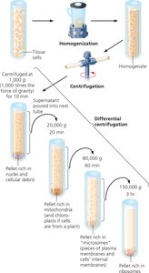
Example: To study protein synthesis, researchers isolate fractions containing ribosomes.
Additional info: Cell fractionation enables bulk preparation of cell components, facilitating biochemical analysis and correlation of structure with function.
Comparing Microscopy Techniques
Different microscopy techniques are suited for specific research purposes:
Light Microscopy: Best for observing living cells and tissues.
SEM: Ideal for surface details and three-dimensional imaging.
TEM: Used for internal cell structure and high-resolution imaging of organelles.
Cryo-EM: Preserves specimens at low temperatures, allowing visualization of structures in their native environment.
Example: To observe the shape changes in a living white blood cell, use a light microscope; for surface texture of a hair, use SEM.
Summary Table: Microscopy Methods
Microscope Type | Purpose | Resolution | Living Cells? |
|---|---|---|---|
Light Microscope | General cell structure, living cells | ~200 nm | Yes |
SEM | Surface details, 3D images | ~1-10 nm | No |
TEM | Internal cell structure | ~1-2 nm | No |
Cryo-EM | Native structure, protein complexes | ~3-5 Å | No |
Key Terms and Definitions
Cytology: Study of cell structure.
Biochemistry: Study of chemical processes within cells.
Organelle: Membrane-bound structure within a cell with a specific function.
Pellet: Material collected at the bottom of a centrifuge tube after spinning.
Supernatant: Liquid above the pellet in a centrifuge tube.
