 Indietro
Microscopy and Diversity of Life: Study Guide for General Biology
Guida di studio - Note intelligenti
 Appunti personalizzati basati sui tuoi materiali, ampliati con definizioni chiave, esempi e contesto.
Microscopy and Diversity of Life
Introduction to Scientific Inquiry and Microscopy
Scientific inquiry in biology involves both controlled experiments and careful observation. Microscopy is a fundamental tool for observing the diversity of life at the cellular level. Proper use of a compound light microscope is essential for accurate scientific observation and documentation.
Scientific Inquiry: Includes experimentation and observation.
Microscopy: Enables visualization of cells and microorganisms.
Lab Safety: Use goggles, gloves, and lab coats when handling stains and living organisms.
Setting Up and Stowing the Compound Light Microscope
Correct setup and maintenance of the microscope ensures accurate results and prevents damage.
Preparation: Remove dust cover, check for damage, clean stage and lenses.
Operation: Turn on light source, follow instructor directions.
Post-Use: Clean lenses, remove slides, store microscope properly.
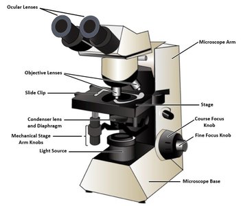
Parts of the Compound Light Microscope
Understanding the function of each microscope component is crucial for effective use.
Ocular Lens: Magnifies the image from the objective lens (usually 10×).
Objective Lenses: Provide varying levels of magnification (e.g., 4×, 10×, 40×, 100×).
Stage: Holds the slide in place.
Coarse and Fine Focus: Adjust the focus for clarity.
Condenser and Diaphragm: Focus and control the amount of light.
Light Source: Illuminates the specimen.
Mechanical Stage: Allows precise movement of the slide.
Microscopy Concepts and Principles
Microscopy relies on several key concepts to produce clear and informative images.
Magnification: Enlargement of the specimen. Total Magnification:
Resolving Power: Ability to distinguish two close objects as separate.
Contrast: Difference between specimen and background, often enhanced by stains.
Depth of Field: Thickness of the sample in focus; decreases with higher magnification.
Field of View: Area visible through the microscope; decreases with higher magnification.
Working Distance: Space between objective lens and slide; decreases with higher magnification.
Cellular Diversity and Classification
Characteristics of Life and Cell Types
All living organisms share fundamental characteristics and are composed of cells, the basic unit of life. Cells are classified as prokaryotic or eukaryotic based on their structure.
Characteristics of Life: Organization, evolutionary adaptation, response to environment, reproduction, regulation, energy processing, growth and development.
Cell Features: Cytosol, cell membrane, DNA, ribosomes.
Prokaryotic Cells: No membrane-bound organelles, DNA in nucleoid, circular chromosomes, plasmids.
Eukaryotic Cells: Membrane-bound organelles, DNA in nucleus, linear chromosomes.
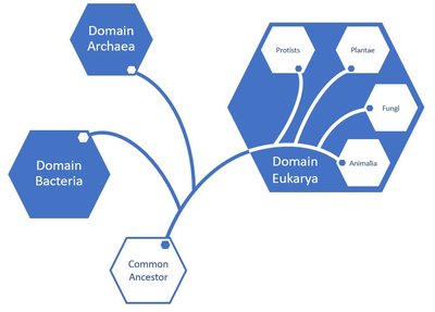
Survey of Eukaryotes
Eukaryotes include protists, fungi, plants, and animals. Each group exhibits unique cellular structures and functions.
Protists
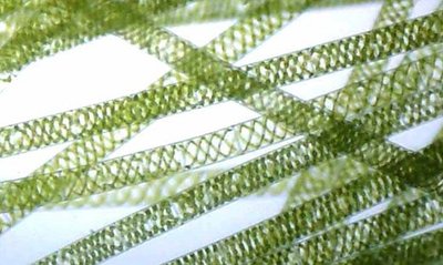
Protists are mostly unicellular eukaryotes, some of which are capable of photosynthesis. Spirogyra crassa is a filamentous algae with spiral chloroplasts.
Observation: Use 4× and 10× objectives to view chloroplasts and explore depth of field.
Field of View: Decreases with higher magnification.
Resolution: Increases with higher magnification.
Depth of Field: Decreases with higher magnification.
Fungi
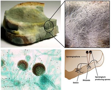
Fungi are multicellular eukaryotic heterotrophs. Rhizopus stolonifer (black bread mold) absorbs nutrients by releasing digestive enzymes.
Hyphae: Filamentous structures that penetrate substrates.
Sporangium: Knob-like structures producing spores.
Staining: Enhances visibility of fungal structures.
Plants
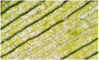
Plants are multicellular, photosynthetic eukaryotes. Elodea is an aquatic plant used to observe cell structure and chloroplasts.
Chloroplasts: Organelles where photosynthesis occurs, containing chlorophyll.
Cell Wall: Provides structural support.
Staining: Iodine is used to enhance visibility of cell structures.
Animals
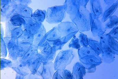
Animals are multicellular eukaryotes. Animal cells lack cell walls and chloroplasts. Human cheek cells are commonly used for observation.
Epithelial Cells: Cells from the lining of the mouth.
Staining: Methylene blue used to highlight cell structures.
Observation: Use 4× and higher objectives, but not 100× oil immersion.
Prokaryotic Survey
Bacterial Morphology and Classification
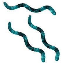
Bacteria are classified by their shape (morphology) and arrangement in colonies.
Bacillus/Bacilli: Rod-shaped
Coccus/Cocci: Spherical
Spirillum/Spirilla: Spiral-shaped
Morphology | Description |
|---|---|
Bacillus | Rod-shaped |
Coccus | Spherical |
Spirillum | Spiral-shaped |


Bacterial Colony Arrangement
Strepto-: Chain-like arrangement
Staphylo-: Grape-like clusters
Arrangement | Description |
|---|---|
Strepto- | Chains |
Staphylo- | Clusters |


Bacterial Staining: Gram Stain Technique
Gram staining differentiates bacteria based on cell wall structure.
Gram-positive: Thick peptidoglycan cell wall, retains purple crystal violet stain.
Gram-negative: Thin peptidoglycan layer, outer phospholipid membrane, stains red with safranin.
Medical Importance: Gram stain status guides antibiotic treatment.
Viruses
Nature and Characteristics of Viruses
Viruses are not cells and are considered neither prokaryotic nor eukaryotic. They are much smaller than cells and require electron microscopes for visualization.
Size: Typically 20–300 nanometers.
Microscopy: Electron microscopes are used to view viruses.
Characteristics of Life: Viruses lack many characteristics of life, such as independent metabolism and reproduction, but can evolve and respond to their environment.
Example: Viruses do not possess cellular structure, cannot reproduce independently, and do not process energy, but they can mutate and adapt.
