 Indietro
Microscopy: Principles, Types, and Applications in Biology
Guida di studio - Note intelligenti
 Appunti personalizzati basati sui tuoi materiali, ampliati con definizioni chiave, esempi e contesto.
Microscopy in Biology
History of the Microscope
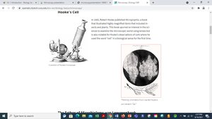
The development of the microscope revolutionized biological sciences by enabling the observation of cells and microorganisms. In 1655, Robert Hooke used a compound microscope to examine cork, coining the term "cells" for the structures he observed. Antonie van Leeuwenhoek was the first to observe single-celled organisms in pond water, further expanding our understanding of microscopic life.
Types of Microscopes
Microscopes are essential tools in biology, allowing scientists to study structures too small to be seen with the naked eye. There are two main types:
Compound Light Microscope: Uses visible light and two sets of lenses to magnify specimens up to 2000x. It is widely used in educational and research settings.
Electron Microscope: Utilizes beams of electrons for much higher magnification (up to 250,000x), enabling observation of viruses, DNA, and cell organelles. There are two types: Scanning Electron Microscope (SEM) and Transmission Electron Microscope (TEM).
Microscope Vocabulary
Understanding key terms is crucial for effective use of microscopes:
Magnification: The increase in apparent size of an object.
Resolution: The ability to distinguish two close points as separate entities; higher resolution reveals finer details.
Field of View: The observable area seen through the microscope.
Depth of Field: The thickness of the specimen that remains in focus at a given magnification.
Magnification Calculations
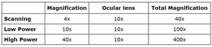
Microscopes typically have multiple objective lenses, each providing different levels of magnification. The total magnification is calculated by multiplying the magnification of the ocular lens (eyepiece) by the objective lens.
Magnification | Ocular lens | Total Magnification | |
|---|---|---|---|
Scanning | 4x | 10x | 40x |
Low Power | 10x | 10x | 100x |
High Power | 40x | 10x | 400x |
For oil immersion (used in microbiology labs):
Objective lens: 100x
Ocular lens: 10x
Total magnification:
Observing the Letter "e" Under the Microscope
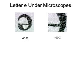
A common introductory exercise in microscopy is observing a printed letter "e" under different magnifications. This helps students understand image orientation, focusing, and the effect of magnification.

As magnification increases, the field of view decreases and finer details become visible. The image may appear inverted or reversed compared to its orientation on the slide.
Depth of Field: Colored Threads Experiment
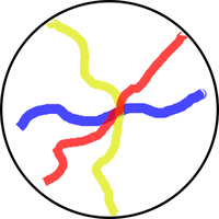
Depth of field refers to the range within a specimen that remains in focus. When examining a slide with colored threads, students observe how only one thread is sharply focused at higher magnifications, while others become blurry. Adjusting the diaphragm to allow less light can increase contrast and detail.
Field of View at Different Magnifications
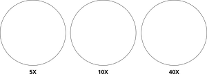
The field of view decreases as magnification increases. This is illustrated by observing the same specimen at scanning, low, and high power objectives.
Examining Cells
Microscopes are fundamental for studying cells, the basic units of life. Prepared slides allow students to observe cell structure, organization, and diversity, reinforcing the importance of microscopy in biological research.
Example: Observing plant or animal cells under high power reveals nuclei, cytoplasm, and other organelles.
Additional info:
Microscopy is directly relevant to General Biology, especially in chapters covering cell structure, function, and scientific inquiry.
Understanding microscope operation and terminology is foundational for laboratory skills in biology.
