 Indietro
Mitosis and Chromosomal Organization: Study Notes for General Biology
Guida di studio - Note intelligenti
 Appunti personalizzati basati sui tuoi materiali, ampliati con definizioni chiave, esempi e contesto.
Mitosis – Cell Division
Genomic Organization
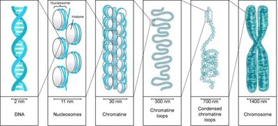
Nuclear DNA in eukaryotes is organized into chromosomes, which are highly condensed structures formed from chromatin during cell division. Chromatin is a complex of DNA and proteins that makes up eukaryotic chromosomes and is typically stringy in appearance when not condensed.
Chromosome: One very long, condensed DNA molecule.
Chromatin: DNA-protein complex; less condensed form present during most of the cell cycle.
Chromosomes form only during cell division for efficient segregation.
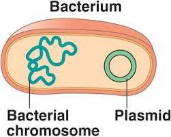
Genomic organization in prokaryotes vs. eukaryotes:
Prokaryotes: One circular chromosome; may have plasmids (small, circular DNA carrying extra genes).
Eukaryotes: Multiple linear chromosomes; each species has a characteristic chromosome number.
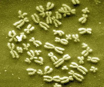
Chromosome numbers: Each organism has a specific number of chromosomes, which does not correlate with complexity. For example, humans have 46 chromosomes, while some ferns have over 1000.
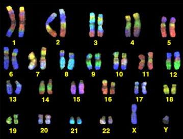
Haploid and Diploid Cells
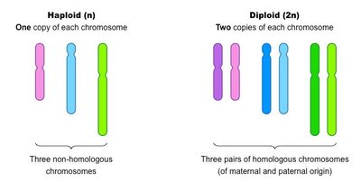
Organisms can have cells with one set (haploid) or two sets (diploid) of chromosomes. In humans, somatic (body) cells are diploid (2n = 46), while gametes (egg and sperm) are haploid (n = 23).
Diploid (2n): Two sets of chromosomes, one from each parent.
Haploid (n): One set of chromosomes, found in gametes.
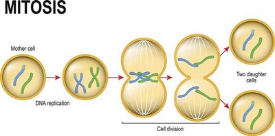
Mitosis produces diploid cells, ensuring genetic consistency in somatic cells.
Chromosome Structure
Single vs. Duplicated Chromosomes
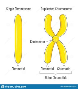
Before cell division, each chromosome is duplicated, forming two sister chromatids joined at a centromere. During mitosis, each chromatid is separated into a new cell.
Chromatid: One half of a duplicated chromosome.
Centromere: Region where sister chromatids are joined.
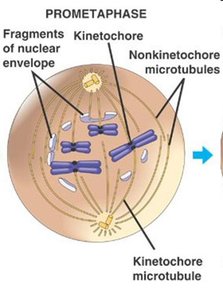
Kinetochore: Protein complex at the centromere for spindle attachment.
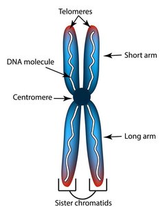
Telomeres are protective caps at chromosome ends, playing a role in aging and stability.
Stages of Mitosis
Overview of Mitosis
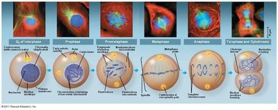
Mitosis is the process by which a diploid cell divides to produce two genetically identical diploid daughter cells. It is essential for growth, repair, and maintenance in multicellular organisms.
Interphase: Cell prepares for division; DNA and organelles are duplicated.
Prophase: Chromosomes condense, nuclear envelope breaks down, spindle forms.
Prometaphase: Spindle fibers attach to kinetochores; chromosomes begin moving.
Metaphase: Chromosomes align at the metaphase plate.
Anaphase: Sister chromatids separate and move to opposite poles.
Telophase: Nuclear envelopes reform, chromosomes decondense.
Cytokinesis: Division of cytoplasm, forming two cells.
Interphase
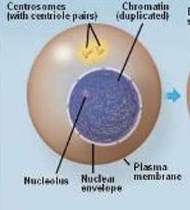
During interphase, the cell grows, duplicates its DNA, and prepares for division. Key checkpoints ensure readiness for mitosis.
G1: Cell growth and organelle duplication
S: DNA replication
G2: Final preparations and checkpoint clearance
Prophase
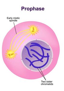
Prophase marks the beginning of mitosis. Chromosomes condense, the nuclear envelope dissolves, and spindle fibers begin to form from centrosomes.
Chromatin condenses into visible chromosomes
Nuclear envelope and nucleolus disappear
Spindle fibers extend and attach to kinetochores
Prometaphase
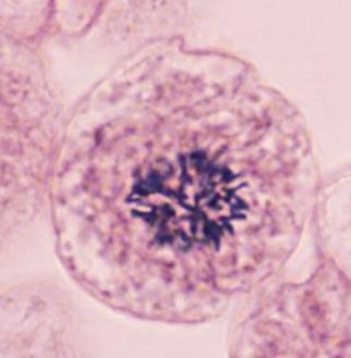
In prometaphase, spindle fibers attach to chromosomes at the kinetochore, and chromosomes begin moving toward the metaphase plate.
Microtubules attach to kinetochores
Nuclear envelope fragments
Metaphase
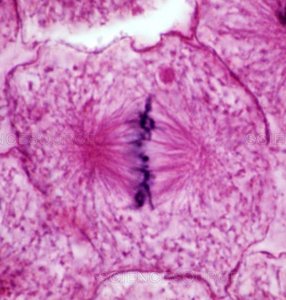
Chromosomes align at the cell's equator, known as the metaphase plate, preparing for separation.
Chromosomes line up single file
Spindle fibers ensure proper alignment
Anaphase
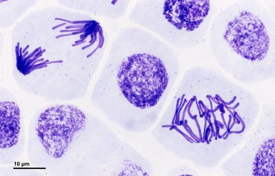
Sister chromatids are pulled apart by spindle fibers and move toward opposite poles of the cell.
Chromatids separate
Each pole receives an identical set of chromosomes
Telophase and Cytokinesis
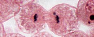
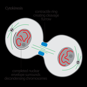
Telophase involves the reformation of nuclear envelopes and decondensation of chromosomes. Cytokinesis divides the cytoplasm, resulting in two daughter cells.
Nuclear envelope reforms
Chromosomes return to chromatin state
Cytokinesis occurs simultaneously or sequentially
Cytokinesis: Animal vs. Plant Cells
Cytokinesis in Animal Cells
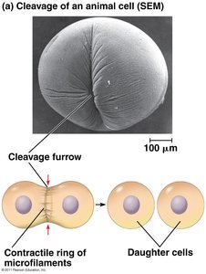
Animal cells undergo cytokinesis via a cleavage furrow, formed by a contractile ring of actin microfilaments and myosin. The furrow deepens until the cell splits into two.
Cleavage furrow forms at metaphase plate
Microfilament ring contracts, pinching cell in two
Cytokinesis in Plant Cells
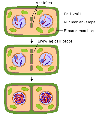
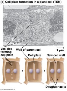
Plant cells cannot undergo cleavage due to their rigid cell wall. Instead, vesicles from the Golgi apparatus form a cell plate at the metaphase plate, which grows and fuses with the plasma membrane, creating a new cell wall.
Vesicles carry cell wall material
Cell plate grows and divides the cell
Summary: Importance of Mitosis
Mitosis produces genetically identical cells, crucial for growth, repair, replacement, immune responses, and maintaining cell size. It ensures that each daughter cell receives a complete set of chromosomes.
Maintains genetic stability
Supports multicellular life
Allows for tissue regeneration and development
Example: Skin cells divide by mitosis to replace lost or damaged cells.
