 Indietro
Mitosis Phase Review: Identifying Cell Types and Structures
Guida di studio - Note intelligenti
 Appunti personalizzati basati sui tuoi materiali, ampliati con definizioni chiave, esempi e contesto.
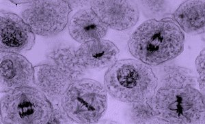
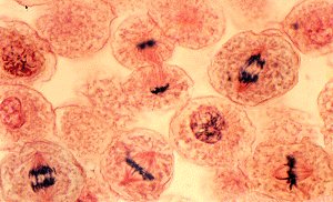
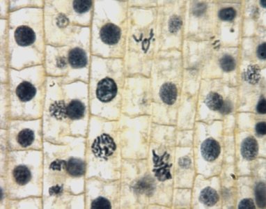
Q1. Identify each set of photos as either ANIMAL or PLANT cells.
Background
Topic: Cell Types and Mitosis
This question tests your ability to distinguish between animal and plant cells based on their appearance under a microscope, especially during mitosis. Recognizing cell types is fundamental in biology because plant and animal cells have distinct features, such as cell walls in plants and cleavage furrows in animals.
Key Terms:
Cell Wall: A rigid structure found in plant cells, absent in animal cells.
Cleavage Furrow: The indentation that begins the process of cytokinesis in animal cells.
Cell Plate: The structure that forms during cytokinesis in plant cells.
Step-by-Step Guidance
Observe the overall shape and arrangement of the cells in each image. Plant cells typically have a more regular, box-like shape due to their cell walls, while animal cells are more rounded.
Look for the presence of a cell wall. If you see a thick, rigid outline around each cell, it is likely a plant cell.
Identify any structures related to cytokinesis. A cleavage furrow indicates animal cells, while a cell plate indicates plant cells.
Compare the three images. Which ones show box-like cells with clear cell walls, and which ones show rounder cells without cell walls?
Try solving on your own before revealing the answer!
Final Answer:
Image 1: Animal cells (rounded, no cell wall visible)
Image 2: Animal cells (rounded, no cell wall visible)
Image 3: Plant cells (box-like shape, cell wall visible)
Images 1 and 2 show animal cells undergoing mitosis, while image 3 shows plant cells. The presence of cell walls in image 3 is a key indicator.
Q2. Label at least one cell for each phase of Mitosis and/or Cytokinesis. (Write the 1st letter of the phase in the cell)
Background
Topic: Phases of Mitosis
This question tests your ability to identify the different phases of mitosis (Prophase, Metaphase, Anaphase, Telophase) and cytokinesis in cells under a microscope. Recognizing these phases is essential for understanding how cells divide and replicate.
Key Terms:
Prophase (P): Chromosomes condense and become visible, nuclear envelope breaks down.
Metaphase (M): Chromosomes align at the cell's equator.
Anaphase (A): Chromosomes are pulled apart toward opposite poles.
Telophase (T): Chromosomes decondense, nuclear envelope reforms.
Cytokinesis (C): Division of the cytoplasm, resulting in two daughter cells.
Step-by-Step Guidance
Examine each cell in the images for visible characteristics: condensed chromosomes, alignment at the equator, separation of chromatids, or formation of new nuclei.
Identify a cell showing condensed chromosomes but no alignment—this is likely Prophase (P).
Find a cell where chromosomes are lined up in the center—this is Metaphase (M).
Look for a cell where chromosomes are being pulled apart—this is Anaphase (A).
Spot a cell where two nuclei are forming—this is Telophase (T).
Identify a cell where the cytoplasm is dividing—this is Cytokinesis (C).
Try solving on your own before revealing the answer!
Final Answer:
P: Cell with condensed chromosomes, nuclear envelope breaking down.
M: Cell with chromosomes aligned at the equator.
A: Cell with chromosomes moving toward opposite poles.
T: Cell with two nuclei forming.
C: Cell showing division of cytoplasm (cleavage furrow or cell plate).
Each phase can be labeled with its first letter in the corresponding cell. Use the images to match these descriptions to actual cells.
Q3. Add the following labels as applicable: Cell Wall, Cell Plate, Nucleus, Nucleolus, Chromosomes, Cleavage Furrow, Spindle Fibers, Equator. (Use each label at least once.)
Background
Topic: Cell Structures During Mitosis
This question tests your knowledge of cellular structures involved in mitosis and cytokinesis. Understanding these structures helps you visualize the mechanics of cell division.
Key Terms:
Cell Wall: Found in plant cells, provides structure.
Cell Plate: Forms during cytokinesis in plant cells.
Nucleus: Contains genetic material.
Nucleolus: Site of ribosome production within the nucleus.
Chromosomes: DNA structures visible during mitosis.
Cleavage Furrow: Indentation in animal cells during cytokinesis.
Spindle Fibers: Structures that move chromosomes during mitosis.
Equator: The central plane where chromosomes align during metaphase.
Step-by-Step Guidance
Look for plant cells (image 3) to label Cell Wall and Cell Plate. The cell wall is the thick outline, and the cell plate appears during cytokinesis.
Identify the Nucleus and Nucleolus in cells not actively dividing; these are usually round and dark within the cell.
Chromosomes are visible as dark, thread-like structures during mitosis.
Cleavage Furrow is seen in animal cells (images 1 and 2) during cytokinesis as an indentation.
Spindle Fibers are visible as lines extending from the poles to the chromosomes during metaphase and anaphase.
The Equator is the central region where chromosomes align during metaphase.
Try solving on your own before revealing the answer!
Final Answer:
Cell Wall: Label in plant cells (image 3).
Cell Plate: Label in plant cells during cytokinesis (image 3).
Nucleus: Label in cells not actively dividing.
Nucleolus: Label within the nucleus.
Chromosomes: Label in cells undergoing mitosis (all images).
Cleavage Furrow: Label in animal cells during cytokinesis (images 1 and 2).
Spindle Fibers: Label in cells during metaphase/anaphase.
Equator: Label in cells during metaphase where chromosomes align.
Each label should be placed in the appropriate cell or structure based on the phase and cell type shown in the images.
