 Indietro
Neurons and Neuronal Signaling: Structure, Function, and Communication
Guida di studio - Note intelligenti
 Appunti personalizzati basati sui tuoi materiali, ampliati con definizioni chiave, esempi e contesto.
The Nervous Tissue
Overview of Nervous Tissue
Nervous tissue is the primary component of the nervous system, responsible for forming a communication network throughout the body. It enables rapid transmission of signals, allowing organisms to sense and respond to their environment.
Main function: Communication and coordination of bodily functions
Cell types: Neurons (excitable cells) and neuroglia (supporting cells)
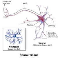
Neurons and Neuroglia
Cell Types in Nervous Tissue
The nervous tissue is composed of two main cell types:
Neurons: Electrically excitable cells, often the longest cells in the body, responsible for transmitting signals
Neuroglia (glia): Supportive cells that maintain the environment around neurons, provide insulation, and supply nutrients
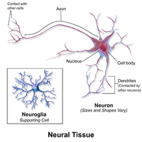
Nervous Tissue at a Cellular Level: Neurons
Structure of a Typical Neuron
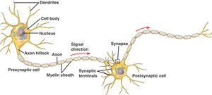
A typical neuron consists of three main parts:
Dendrite: Branched extensions that receive signals from other neurons
Soma (cell body): Contains the nucleus and most organelles
Axon: Long projection that transmits signals to other cells
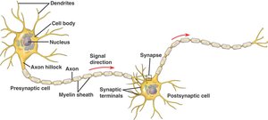
The Dendrite
Dendrites are specialized for receiving electrochemical signals from other neurons and transmitting this information to the soma.
Function: Input region of the neuron
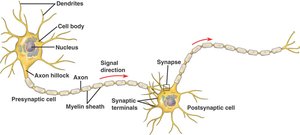
The Soma
The soma is the cell body of the neuron, housing the nucleus and most organelles.
Function: Integrates incoming signals and maintains cell health
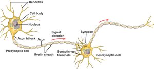
The Axon
The axon is a long projection that carries electrical or chemical signals to the synapse.
Myelin sheath: Insulates the axon, increasing signal speed
Neurons and Action Potentials
Membrane Potential
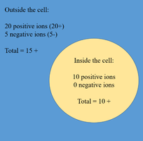
The membrane potential is the difference in electric potential between the inside and outside of a cell, determined by:
Charge difference: Distribution of positive and negative ions
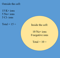
Ion concentration difference: Created by ion channels and pumps
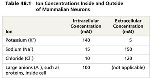
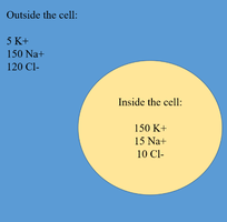
Concentration Differences in a Neuron
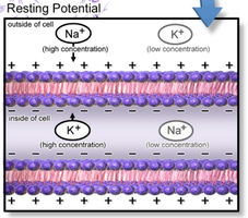
Neurons maintain specific ion concentrations inside and outside the cell.
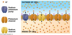
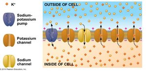
Sodium-potassium pump: Uses ATP to maintain gradients
Ion and Charge Gradient
Ion gradients are maintained because ions cannot freely diffuse across the membrane.
Ion channels: Allow selective movement of ions
The Nernst Equation
The equilibrium potential for each ion can be calculated using the Nernst equation:
Formula:
Equilibrium potential: The voltage at which there is no net movement of the ion
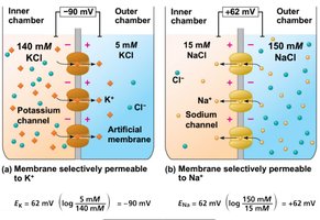
The Actual Membrane Potential
The actual membrane potential depends on the permeability of the membrane to different ions.
More K+ movement: Potential approaches -90 mV
More Na+ movement: Potential approaches +62 mV

The Neuron’s Resting Membrane Potential
At rest, the neuron’s membrane potential is typically between -60 and -80 mV.
Resting potential: Maintained by ion gradients and selective permeability
Changes in Polarization
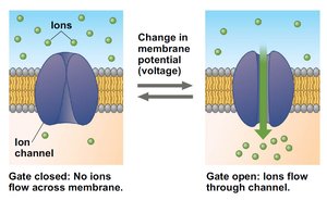
Voltage-gated ion channels open or close in response to stimuli, altering membrane permeability and potential.
Hyperpolarization: Opening K+ channels makes the membrane more negative
Depolarization: Opening Na+ channels makes the membrane more positive
Graded Potentials and Action Potentials
External stimuli trigger changes in membrane potential.
Graded potentials: Magnitude varies with stimulus strength
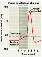
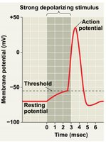
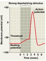
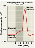
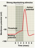
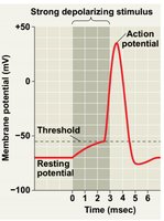
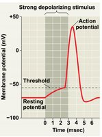
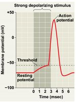
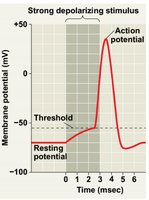
Action potentials: Occur if membrane potential reaches threshold (~-50 mV)
Stages of the Action Potential
The action potential consists of several stages:
Resting potential: Most channels closed, ~-70 mV
Threshold: Depolarization to ~-50 mV triggers action potential
Rising phase: Na+ channels open, influx of Na+, membrane potential increases
Peak: Na+ channels inactivate, K+ channels open
Falling phase: K+ efflux, membrane potential decreases
Undershoot: Membrane potential approaches -90 mV, then returns to rest
Refractory period: Na+ channels temporarily inactivated, prevents backward propagation
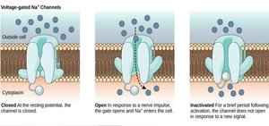
Action Potentials Move in One Direction
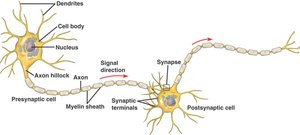
Action potentials are generated at the axon hillock and travel toward the synaptic terminals.
Unidirectional movement: Inactivated Na+ channels prevent backward propagation
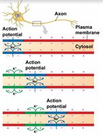
Adaptations to Action Potential Speed
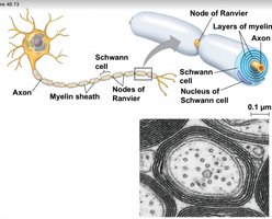
The speed of action potential transmission increases with axon diameter and myelin sheath insulation.
Myelin sheath: Insulates axons, increases speed via saltatory conduction
Neuron Communication
Synapses
Neurons communicate at synapses, where two neurons are in close proximity.
Electrical synapses: Direct current flow via gap junctions
Chemical synapses: Neurotransmitters carry information across the synaptic cleft
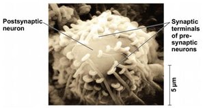
The Chemical Synapse
When an action potential reaches the axon terminal, neurotransmitters are released.
Synapse components: Axon terminal, synaptic cleft, postsynaptic dendrite or soma
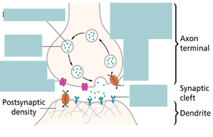
Neurotransmitter Release and Reception
Neurotransmitters are released from the presynaptic neuron by exocytosis, cross the synaptic cleft, and bind to receptors on the postsynaptic neuron.
Receptors: Ligand-gated ion channels (fast) or signaling receptors (slow, long-lasting effects)
Postsynaptic Potentials
Binding of neurotransmitters can trigger postsynaptic potential changes:
Excitatory postsynaptic potentials (EPSPs): Depolarizations toward threshold
Inhibitory postsynaptic potentials (IPSPs): Hyperpolarizations away from threshold
Summation of EPSPs and IPSPs
Multiple EPSPs and IPSPs can be combined:
Temporal summation: Rapid succession from a single synapse
Spatial summation: Simultaneous input from multiple synapses
Neurotransmitter Clearance
Neurotransmitter action ceases when they are removed from the synaptic cleft by diffusion, enzymatic inactivation, or recapture.
Types of Neurotransmitters
Neurotransmitters are classified into four groups:
Amino acids: e.g., glycine
Biogenic amines: e.g., epinephrine, dopamine, serotonin
Neuropeptides: e.g., endorphins
Gases: e.g., nitric oxide (NO)
The Central Nervous System
Structure of the CNS
The central nervous system consists of the brain and spinal cord.
Function: Integration and processing of information
The Blood Brain Barrier (BBB)
The BBB protects the brain by preventing most molecules from crossing blood vessels into brain tissue.
Structure: Tight junctions between supporting cells
Function: Prevents infection, but also blocks many drugs
Alzheimer’s Disease and the BBB
Alzheimer’s disease is characterized by amyloid-β peptide deposits (plaques).
Therapies: Target amyloid creation/removal, but BBB limits drug effectiveness
Ultrasound and BBB Opening
Ultrasound can temporarily open the BBB, allowing drugs or molecules to enter the brain.
Experimental results: Reduction of plaque and improved memory in mice
Attendance Questions
Potassium Ion Channels Open, Sodium Ion Channels Closed
Answer: Hyperpolarized
Explanation: Opening K+ channels increases K+ efflux, making the inside more negative
2 EPSPs and 2 IPSPs
Answer: Not undergo an action potential
Explanation: IPSPs counteract EPSPs, so the combined effect does not reach threshold
