Indietro
BIO 102 Chapter 42 Part 1 B
Guida di studio - Note intelligenti
 Appunti personalizzati basati sui tuoi materiali, ampliati con definizioni chiave, esempi e contesto.
Relationship Between Structure and Function
The vertebrate circulatory system relies on blood vessels whose structure closely matches their function. This relationship is fundamental to understanding how blood pressure and flow are regulated throughout the body.
Blood Vessel Types and Functions
Major Blood Vessel Classes
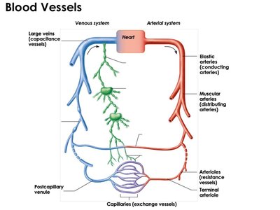
Blood vessels are responsible for transporting blood, regulating blood flow and pressure, and facilitating exchange of substances. The three main types of blood vessels are:
Arteries: Carry blood away from the heart (efferent), act as distribution vessels.
Capillaries: Serve as exchange vessels, allowing for the transfer of nutrients, gases, and waste.
Veins: Return blood to the heart (afferent), act as collection and drainage vessels.
Arteries are further subdivided based on their size and function:
Elastic (Conducting) Arteries: Large vessels like the aorta and pulmonary trunk; act as pressure reservoirs, expand and recoil to maintain continuous blood flow.
Muscular (Distributing) Arteries: Medium-sized arteries (e.g., femoral artery); direct blood to specific regions.
Arterioles: Smallest arteries; regulate blood flow to capillaries and control resistance.
Veins and venules collect blood from capillary beds and return it to the heart. Large veins serve as blood reservoirs and operate under low pressure.
Blood Vessel Structure and Function
Layers of Blood Vessels
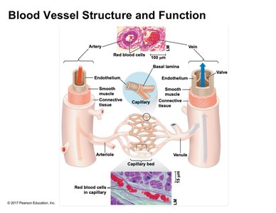
Arteries and veins share a similar basic structure, but differ in the distribution of their components:
Endothelium: Innermost layer, continuous with the heart's endocardium; provides a smooth, friction-reducing surface.
Smooth Muscle: Controls vessel diameter and blood flow; more prominent in arteries.
Connective Tissue: Outermost layer; provides support and prevents overstretching, especially important in arteries.
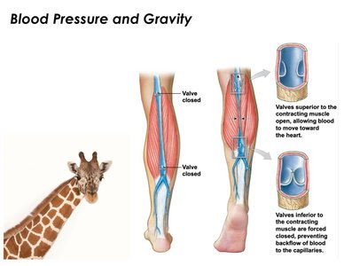
Capillaries consist mainly of endothelial cells and basal lamina, allowing for single-file passage of red blood cells and efficient exchange. Veins contain valves to prevent backflow, especially in the legs, counteracting gravity.
Hemodynamics: Blood Flow, Velocity, and Pressure
Changes Across Circulatory System
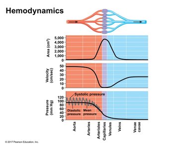
Blood flow characteristics change as blood moves through different vessels:
Cross-sectional Area: Increases dramatically in capillaries due to extensive branching.
Velocity: Inversely related to cross-sectional area; fastest in the aorta, slowest in capillaries, increases again in veins.
Pressure: Highest in arteries (especially aorta), decreases through capillaries and veins. Pressure is pulsatile in arteries due to systolic and diastolic cycles.
Mean arterial pressure is used as a quantitative measure for tissue perfusion.
Regulation of Blood Pressure
Vasoconstriction and Vasodilation
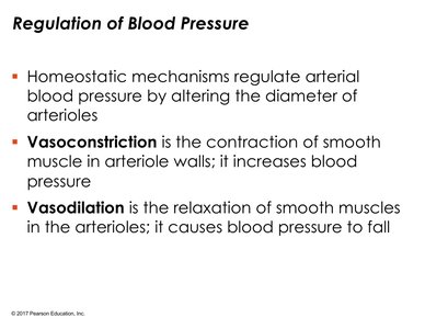
Blood pressure is regulated by altering the diameter of arterioles:
Vasoconstriction: Contraction of smooth muscle in arteriole walls increases blood pressure.
Vasodilation: Relaxation of smooth muscle decreases blood pressure.
These mechanisms are essential for maintaining homeostasis and responding to physiological demands.
Cell Signaling in Blood Vessel Regulation
Smooth Muscle Relaxation Pathway
Cell communication and signal transduction play a role in regulating blood vessel diameter:
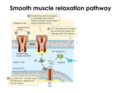
Acetylcholine binds to receptors on endothelial cells, activating the receptor and producing IP3 (a second messenger).
IP3 binds to ligand-gated channels on the endoplasmic reticulum, releasing Ca2+ into the cytosol.
Ca2+ activates nitric oxide synthase, producing nitric oxide (NO) from arginine.
Nitric Oxide and Muscle Relaxation
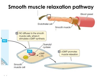
Nitric oxide diffuses to smooth muscle cells, stimulating cyclic GMP synthesis via guanylyl cyclase. Cyclic GMP promotes muscle relaxation, increasing blood flow. Drugs like nitroglycerin and sildenafil (Viagra) act on this pathway to enhance blood flow.
Blood Pressure and Gravity
Venous Return and Adaptations
Blood pressure decreases in veins, making venous return to the heart challenging, especially against gravity. Mechanisms such as the muscular pump (skeletal muscle contraction) and venous valves assist in returning blood to the heart. Animals with long necks, like giraffes, have adaptations for high systolic pressure to ensure blood reaches the head.
Measuring Blood Pressure
Sphygmomanometer Use
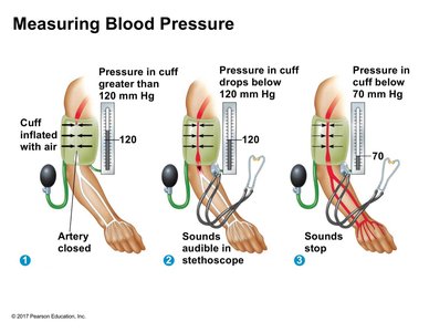
Blood pressure is measured using a sphygmomanometer (blood pressure cuff) on the brachial artery:
Cuff inflated above systolic pressure (>120 mm Hg): artery closed, no sounds.
Cuff pressure drops below systolic: sounds audible, indicating systolic pressure.
Cuff pressure drops below diastolic (<70 mm Hg): sounds stop, indicating diastolic pressure.
Systolic and diastolic pressures are key indicators of circulatory efficiency.
Capillary Function and Microcirculation
Capillary Beds and Sphincters
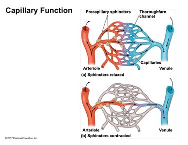
Capillaries form networks called capillary beds, facilitating microcirculation between arterioles and venules. Precapillary sphincters regulate blood flow into capillaries, allowing selective perfusion based on local chemical conditions and physiological needs (e.g., fight or flight response).
Fluid Exchange Between Capillaries and Interstitial Fluid
Blood Pressure and Osmotic Pressure
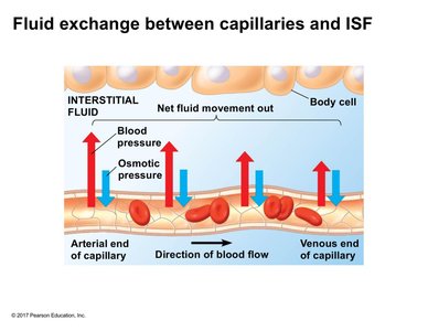
Fluid exchange occurs primarily at the arteriole end of capillaries due to higher blood pressure. Plasma proteins create osmotic pressure, drawing water back into capillaries. The balance between blood pressure and osmotic pressure determines net fluid movement. Excessive fluid loss can lead to edema, commonly seen in pregnancy due to increased blood volume and venous compression.
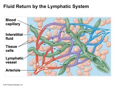
Fluid Return by the Lymphatic System
Lymphatic System Role
The lymphatic system returns excess fluid lost from capillaries to circulation. Lymphatic vessels are highly permeable, allowing tissue fluid to drain into them. Lymph moves through one-way valves and eventually drains into subclavian veins. Unlike the cardiovascular system, the lymphatic system lacks a muscular pump and relies on skeletal muscle contraction for fluid movement.