 Indietro
Polymerase Chain Reaction (PCR) and Genetic Testing: Application to Polycystic Kidney Disease
Guida di studio - Note intelligenti
 Appunti personalizzati basati sui tuoi materiali, ampliati con definizioni chiave, esempi e contesto.
PCR and Genetic Testing in Polycystic Kidney Disease
Introduction to Polycystic Kidney Disease (PKD)
Polycystic kidney disease (PKD) is a common inherited disorder that leads to the formation of multiple cysts in the kidneys, often resulting in kidney failure. Genetic testing is crucial for confirming a diagnosis and identifying mutations in genes such as PKD1 or PKD2.
Polycystic kidney disease (PKD): A genetic disorder characterized by the growth of numerous cysts in the kidneys.
Cyst: A closed, sac-like structure filled with fluid, air, or other material, which can form in various body tissues.
Genetic testing: Used to detect mutations in specific genes associated with PKD.
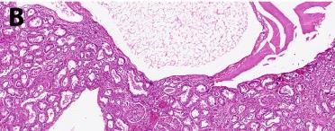
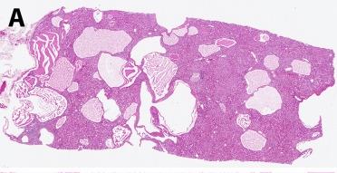
Histological Features of PKD
Histological analysis of kidney tissue from PKD patients reveals numerous cysts of varying sizes, disrupting normal kidney architecture.
Hematoxylin and eosin staining: Commonly used to visualize tissue structure and identify cysts in kidney biopsies.
Polymerase Chain Reaction (PCR): Principles and Application
Overview of PCR
The polymerase chain reaction (PCR) is a laboratory technique used to amplify specific DNA sequences, making millions of copies from a small initial sample. PCR is essential for genetic testing, including the detection of mutations in the PKD2 gene.
Amplification: The process of making many copies of a specific DNA region.
Applications: Mutation detection, genetic diagnosis, forensic analysis, and research.
Requirements for PCR
To perform PCR, several key components are required:
Template DNA: Genomic DNA containing the target gene (e.g., PKD2).
DNA primers: Short, single-stranded DNA sequences that flank the target region and provide a starting point for DNA synthesis.
DNA nucleotides (dNTPs): Building blocks for new DNA strand synthesis.
Heat-stable DNA polymerase: Enzyme (e.g., Taq polymerase) that synthesizes new DNA strands at high temperatures.
PCR Steps and Mechanism
PCR consists of repeated cycles, each with three main steps:
Denaturation (95°C): Double-stranded DNA is separated into single strands by heat.
Annealing (50–65°C): Primers bind (anneal) to their complementary sequences on the single-stranded DNA.
Extension (72°C): DNA polymerase extends the primers, synthesizing new DNA strands.
After multiple cycles, the target DNA region is exponentially amplified.
Designing PCR Primers
Primers must be designed to bind to the ends of the target DNA region, with their 3' ends oriented toward each other to allow amplification of the intervening sequence.
Forward primer: Binds to the 3' end of the template strand.
Reverse primer: Binds to the 3' end of the complementary strand.
Primer orientation: DNA polymerase adds nucleotides to the 3' end of each primer.
Example: Primer Design for PKD2 Gene
Given the following DNA sequence:
5’ GCTGCCGGGAC...TGGAAACCATCA… 3’ 3’ CGACGGCCCTG...ACCTTTGGTAGT… 5’
Primers that would amplify the entire target region are:
Forward primer: 5’ GCTGCC 3’
Reverse primer: 5’ GGTAGT 3’ (complementary to the 3' end of the bottom strand)
Primers must face each other to allow amplification of the intervening sequence.
Key Enzyme: Taq Polymerase
Taq polymerase is a heat-stable DNA polymerase isolated from the thermophilic bacterium Thermus aquaticus, found in hot springs. Its stability at high temperatures makes it ideal for PCR.
Thermostability: Taq polymerase remains active after repeated heating cycles.
Source: Thermus aquaticus (Yellowstone hot springs).

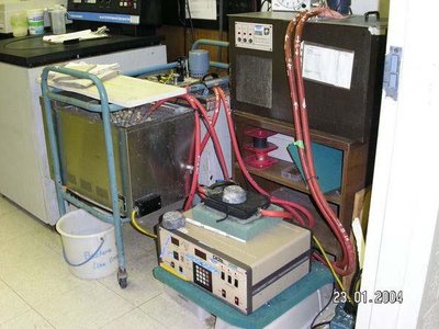
PCR Machines: Past and Present
PCR requires precise temperature cycling, achieved using a thermocycler. Early machines were large and manually operated, while modern thermocyclers are compact and automated.
Early PCR machines: Used water baths at different temperatures.
Modern thermocyclers: Automated, programmable devices for rapid cycling.

Genetic Analysis and Mutation Detection
Sequencing and Mutation Identification
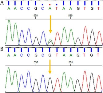
After PCR amplification, DNA sequencing can reveal mutations in the target gene. In PKD, sequencing can distinguish between wild-type and mutant alleles.
Heterozygous mutation: Presence of both wild-type and mutant peaks in sequencing chromatograms (e.g., G/A genotype).
Dominant mutation: A single mutant allele can cause disease if the mutation affects gene function.
Function of PKD2 Gene Product
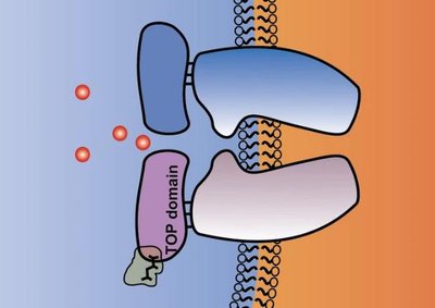
The PKD2 gene encodes polycystin-2, a protein that forms a channel in the cell membrane of kidney cells. Together with polycystin-1, it regulates calcium ion transport, cell growth, and cell signaling.
Polycystin-2: Channel protein for calcium ions.
Role in cell signaling: Calcium influx triggers intracellular signaling cascades.
Regulation: Polycystin-1 and polycystin-2 interact to control cell proliferation and migration.
Comparison: PCR vs. In Vivo DNA Replication
Key Differences and Similarities
PCR and in vivo DNA replication share fundamental principles but differ in their mechanisms and components. The following table summarizes the main features:
Feature | In Vivo Replication | PCR |
|---|---|---|
Strand Separation | Helicase enzyme | Heat |
Enzyme for Elongation | DNA polymerase | Taq or other thermostable DNA polymerase |
Primers | RNA | DNA |
Ligase Needed? | Yes, to join Okazaki fragments | No, no lagging strand |
Similarities | Both require nucleotides, a DNA template, and synthesize new DNA in the 5'→3' direction | |
Summary
PCR is a powerful tool for amplifying specific DNA sequences, enabling genetic testing for diseases such as polycystic kidney disease. Understanding the principles of PCR, primer design, and the comparison with natural DNA replication is essential for modern molecular biology and genetics.
