 Indietro
Protein Structure and Function: Study Notes for General Biology
Guida di studio - Note intelligenti
 Appunti personalizzati basati sui tuoi materiali, ampliati con definizioni chiave, esempi e contesto.
Protein Structure
Amino Acid Structure
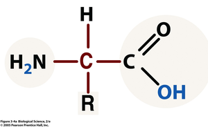
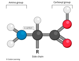
Amino acids are the fundamental building blocks of proteins. Each amino acid consists of a central carbon atom (α-carbon) bonded to four groups: an amino group, a carboxyl group, a hydrogen atom, and a variable side chain (R group). The properties of the side chain determine the chemical nature of the amino acid.
Central Carbon (α-carbon): The core atom to which all groups are attached.
Amino Group (–NH2): Positively charged at neutral pH.
Carboxyl Group (–COOH): Negatively charged at neutral pH.
Side Chain (R group): Determines the identity and properties of the amino acid.
Classification of Amino Acids by Side Chain Properties
Amino acids are classified based on the chemical nature of their side chains:
Nonpolar: Side chains contain mostly C and H (sometimes S); hydrophobic.
Polar Uncharged: Side chains have partial charges (d+ or d–); hydrophilic.
Polar Charged: Side chains have full positive or negative charges; hydrophilic.
Example: Amino acids with O-H or charged groups in their side chains are hydrophilic, while those with only C, H, and S are hydrophobic.
Polymerization of Amino Acids
Proteins are polymers formed by linking amino acid monomers through dehydration (condensation) reactions, which remove water and form peptide bonds.
Dehydration (Condensation) Reaction: Joins amino acids by removing H2O.
Peptide Bond: Covalent bond between the carboxyl group of one amino acid and the amino group of another.
Hydrolysis Reaction: Breaks peptide bonds by adding water.
Equation: 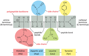
Levels of Protein Structure
Primary Structure
The primary structure is the linear sequence of amino acids in a polypeptide chain, held together by peptide bonds. This sequence determines all higher levels of structure and ultimately the protein's function.
Peptide Bonds: Covalent bonds linking amino acids.
Sequence: Unique order of amino acids.

Secondary Structure
Secondary structure refers to local folding patterns within the polypeptide, stabilized by hydrogen bonds between backbone atoms. The two main types are:
α-Helix: Coiled structure stabilized by hydrogen bonds.
β-Pleated Sheet: Sheet-like structure formed by hydrogen bonds between parallel strands.
Example: Hydrogen bonds within the backbone produce α-helices and β-sheets.
Tertiary Structure
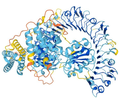
Tertiary structure is the overall three-dimensional shape of a single polypeptide, determined by interactions among side chains (R groups). Four main types of interactions stabilize tertiary structure:
Hydrophobic Interactions: Nonpolar side chains cluster away from water.
Hydrogen Bonds: Between polar side chains.
Disulfide Bridges: Covalent bonds between cysteine residues.
Ionic Bonds: Between charged side chains.
Quaternary Structure
Quaternary structure arises when two or more polypeptide subunits interact to form a functional protein complex. The arrangement and interaction of these subunits are essential for the protein's activity.
Subunit Interaction: Multiple polypeptides associate.
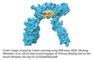
Functional Complex: Example: Hemoglobin, insulin receptor.
Summary Table: Levels of Protein Structure
Level | Description | Bonds/Interactions |
|---|---|---|
Primary | Sequence of amino acids | Peptide bonds |
Secondary | Local folding (α-helix, β-sheet) | Hydrogen bonds |
Tertiary | 3D shape of polypeptide | Hydrophobic, hydrogen, disulfide, ionic |
Quaternary | Association of subunits | Same as tertiary, between subunits |
Protein Folding and Chaperones
Role of Chaperone Proteins
Chaperone proteins assist in the proper folding of other proteins, preventing misfolding and aggregation. Proper folding is essential for protein function, as structure determines function.
Chaperones: Facilitate correct folding.
Misfolded Proteins: Can lead to malfunction and disease.
Example: Insulin must be properly folded to regulate blood sugar; misfolded insulin leads to diabetes.
Structure Determines Function
Consequences of Altered Primary Structure
A change in the primary structure (amino acid sequence) can alter protein folding, leading to loss of function or disease. For example, a mutation in insulin's sequence can cause improper folding and unregulated blood sugar levels.
Mutation: Alters amino acid sequence.
Misfolding: Results in nonfunctional protein.
Disease Example: Diabetes due to misfolded insulin.
Key Terms and Definitions
Amino Acid: Organic molecule with amino, carboxyl, hydrogen, and side chain groups.
Peptide Bond: Covalent bond joining amino acids.
Polypeptide: Chain of amino acids.
Protein: Folded polypeptide with specific function.
Chaperone: Protein assisting in folding.
Summary
Proteins are essential biomolecules with diverse functions, determined by their unique structures. Understanding the hierarchical levels of protein structure and the chemical properties of amino acids is fundamental to grasping how proteins work in living cells.
