 Indietro
Protein Structure and Function: Study Notes for General Biology
Guida di studio - Note intelligenti
 Appunti personalizzati basati sui tuoi materiali, ampliati con definizioni chiave, esempi e contesto.
Protein Structure and Function
Introduction to Proteins
Proteins are the most abundant and versatile macromolecules in living organisms. They are essential for nearly every biological process, serving as catalysts, structural components, signaling molecules, and more. Understanding protein structure is fundamental to understanding their diverse functions in cells.
Amino acids are the building blocks of proteins.
Proteins are polymers composed of 20 different amino acids, each with a unique side chain (R-group).
The sequence and chemical properties of amino acids determine protein structure and function.
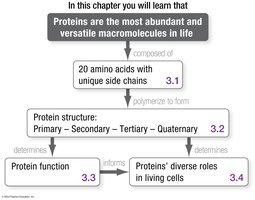
3.1 Amino Acids and Their Polymerization
Structure of Amino Acids
All amino acids share a common core structure, consisting of a central carbon atom (the alpha carbon) bonded to four different groups:
A hydrogen atom (H)
An amino group (–NH2)
A carboxyl group (–COOH)
A variable side chain, known as the R-group
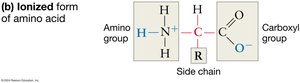
In aqueous solutions, amino and carboxyl groups ionize, which helps amino acids stay in solution and affects their chemical reactivity.
The Nature of Side Chains (R-Groups)
The R-group, or side chain, is what makes each of the 20 amino acids unique. The properties of amino acids vary because their R-groups differ in size, shape, charge, hydrophobicity, and reactivity.
Some R-groups contain functional groups that can participate in chemical reactions.
Others are composed solely of carbon and hydrogen, making them nonreactive and hydrophobic.
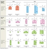
Polarity and Charge of R-Groups Affect Solubility
The chemical nature of the R-group determines whether an amino acid is hydrophilic (water-loving) or hydrophobic (water-fearing):
Charged and polar R-groups interact with water and are hydrophilic.
Nonpolar R-groups do not interact with water and are hydrophobic.
Classification of amino acids based on R-group:
Charged (acidic or basic)
Uncharged polar
Nonpolar
To classify an amino acid, ask:
Does the side chain have a negative charge? (Acidic)
Does the side chain have a positive charge? (Basic)
If uncharged, does it contain an oxygen atom? (Uncharged polar)
If none of the above, it is nonpolar.
Polymerization: Formation of Peptide Bonds
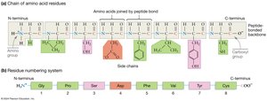
Amino acids link together via peptide bonds, which form between the carboxyl group of one amino acid and the amino group of another through a condensation reaction (releasing water).

The resulting chain of amino acids is called a polypeptide. The sequence of amino acids in a polypeptide is called its primary structure.
The Peptide-Bonded Backbone
R-group orientation: Side chains extend out from the backbone, allowing interactions with each other and the environment.
Directionality: Polypeptides have an N-terminus (free amino group) and a C-terminus (free carboxyl group). By convention, sequences are written from N- to C-terminus.
Flexibility: The peptide bond itself is rigid, but single bonds on either side allow rotation, giving the polypeptide flexibility.
Oligopeptides, Polypeptides, and Proteins
Oligopeptide (peptide): Fewer than 50 amino acids.
Polypeptide: More than 50 amino acids.
Protein: A complete, functional form of a polypeptide chain.
3.2 What Do Proteins Look Like?
Levels of Protein Structure
Protein structure is hierarchical, with four levels:
Primary structure: Unique sequence of amino acids.
Secondary structure: Local folding patterns (e.g., alpha-helices and beta-pleated sheets) stabilized by hydrogen bonds between backbone atoms.
Tertiary structure: Overall three-dimensional shape formed by interactions among R-groups (hydrogen bonds, hydrophobic interactions, van der Waals forces, covalent disulfide bonds, ionic bonds).
Quaternary structure: Association of multiple polypeptide subunits into a functional protein complex.
Each level of structure is critical for the protein's final shape and function.
Primary Structure
Determined by the sequence of amino acids.
Even a single amino acid change can drastically alter protein function (e.g., sickle cell hemoglobin).
Secondary Structure
Formed by hydrogen bonds between the carbonyl group of one amino acid and the amino group of another.
Common types: Alpha-helix and beta-pleated sheet.
Tertiary Structure
Results from interactions among R-groups and between R-groups and the backbone.
Types of interactions: hydrogen bonds, hydrophobic interactions, van der Waals forces, covalent disulfide bonds, ionic bonds.
Determines the overall 3D shape of the protein.
Quaternary Structure
Formed when two or more polypeptide chains (subunits) interact to form a functional protein.
Examples: Dimers (two subunits), homodimers (identical subunits), heterodimers (different subunits), and large macromolecular complexes (e.g., ribosomes).
Level of Structure | Description | Stabilizing Bonds/Interactions |
|---|---|---|
Primary | Sequence of amino acids | Peptide bonds |
Secondary | Alpha-helix, beta-sheet | Hydrogen bonds (backbone) |
Tertiary | 3D folding of a single polypeptide | R-group interactions (hydrogen, ionic, hydrophobic, van der Waals, disulfide) |
Quaternary | Association of multiple polypeptides | Same as tertiary (between subunits) |
3.3 Folding and Function
Protein Folding
Proper folding is essential for protein function. Folding is often spontaneous, driven by chemical bonds and interactions that make the folded state more stable (lower potential energy) than the unfolded state.
Denatured proteins (unfolded) lose their function.
Molecular chaperones are proteins that assist in the proper folding of other proteins, preventing inappropriate interactions and aggregation.
Protein folding can be regulated and may require binding to other molecules to achieve the final functional form.
Protein Misfolding and Prions
Misfolded proteins can be "infectious," causing normal proteins to adopt abnormal conformations. Prions are misfolded proteins that can induce other proteins to misfold, leading to diseases such as mad cow disease.
3.4 Protein Functions Are as Diverse as Protein Structures
Major Functions of Proteins
Catalysis: Enzymes speed up chemical reactions by lowering activation energy.
Structure: Provide support and shape to cells and tissues (e.g., collagen, keratin).
Movement: Motor proteins move cells or molecules within cells (e.g., actin, myosin).
Signaling: Transmit signals between cells (e.g., hormones, receptors).
Transport: Carry molecules across membranes or throughout the body (e.g., hemoglobin).
Defense: Antibodies protect against pathogens.
Enzymes as Catalysts
Enzymes are proteins that function as biological catalysts. They bind reactants (substrates) at a specific location called the active site, holding them in an orientation that makes the reaction more likely to occur.
Proteins and the Origin of Life
While amino acids and small peptides can form under prebiotic conditions, it remains unclear if proteins were the first self-replicating molecules. Proteins would need to replicate, evolve, and carry information to serve as the origin of life. Nucleic acids are now considered more likely candidates for the first informational molecules.
