 Indietro
Signal Transduction and Cell Signaling in Animal Cells
Guida di studio - Note intelligenti
 Appunti personalizzati basati sui tuoi materiali, ampliati con definizioni chiave, esempi e contesto.
Signal Transduction
Overview of Signal Transduction
Signal transduction is the process by which a cell converts an external signal into a functional response. This involves a series of molecular events, typically initiated by the binding of a signaling molecule (ligand) to a receptor, leading to changes inside the cell. - Key Point 1: Signal transduction allows cells to respond to their environment and communicate with other cells. - Key Point 2: The process often involves amplification, where a small signal leads to a large cellular response.
Main Types of Receptors in Animal Cells
Animal cells utilize several types of receptors to detect and respond to signals: - G protein-coupled receptors (GPCRs): Mediate responses to hormones, neurotransmitters, and environmental stimuli. - Receptor tyrosine kinases (RTKs): Involved in growth, differentiation, and metabolism. - Ion channel receptors: Control ion flow across membranes, crucial for nerve and muscle function. - Intracellular receptors: Bind molecules that can cross the membrane, such as steroid hormones.
G Protein-Coupled Receptors (GPCRs)
GPCR Signaling: Activation
GPCRs are membrane proteins that activate G proteins upon ligand binding. The G protein exchanges GDP for GTP, becoming active and triggering downstream effects. - Key Point 1: GPCRs are the largest family of cell surface receptors. - Key Point 2: Activation involves conformational changes and nucleotide exchange. 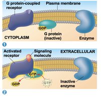
GPCR Signaling: Cellular Response and Hydrolysis
The activated G protein interacts with target enzymes, leading to a cellular response. GTP is hydrolyzed to GDP, inactivating the G protein. - Key Point 1: Cellular responses can include changes in metabolism, gene expression, or ion channel activity. - Key Point 2: GTP hydrolysis ensures the signal is transient. 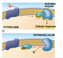
Second Messengers and Signal Amplification
Second messengers such as cyclic AMP (cAMP) and Ca2+ amplify the signal inside the cell. - Key Point 1: Second messengers are small molecules that propagate the signal. - Key Point 2: Amplification allows a single ligand to produce a large response. 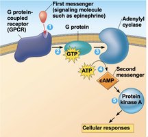
Example: Glycogen Breakdown by Epinephrine
Epinephrine binding to GPCRs triggers a cascade that amplifies the signal, resulting in rapid glycogen breakdown. 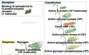 Example: In muscle cells, epinephrine leads to the production of glucose-1-phosphate from glycogen, fueling the 'fight or flight' response.
Receptor Tyrosine Kinases (RTKs)
RTK Structure and Activation
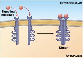
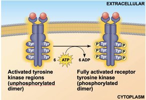
RTKs are transmembrane proteins with intrinsic kinase activity. Ligand binding induces dimerization and autophosphorylation. - Key Point 1: RTKs phosphorylate tyrosine residues on themselves and other proteins. - Key Point 2: Dimerization is essential for activation. 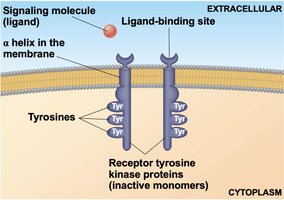
RTK Signaling: Cellular Response
Phosphorylated RTKs recruit and activate relay proteins, leading to multiple cellular responses. - Key Point 1: RTK signaling regulates cell growth, division, and differentiation. - Key Point 2: Abnormal RTK activity is linked to cancer. 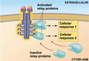
RTKs and Cancer
Dysregulation of RTKs can drive tumor growth and survival. Many anti-cancer drugs target RTKs to inhibit aberrant signaling. 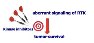
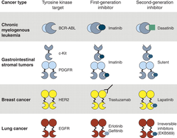
Table: Cancer Types and RTK Inhibitors
Cancer Type | Tyrosine Kinase Target | First-generation Inhibitor | Second-generation Inhibitor |
|---|---|---|---|
Chronic myelogenous leukemia | BCR-ABL | Imatinib | Dasatinib |
Gastrointestinal stromal tumors | c-Kit, PDGFR | Imatinib | Sutent |
Breast cancer | HER2 | Trastuzumab | Lapatinib |
Lung cancer | EGFR | Erlotinib, Gefitinib | Irreversible inhibitors (e.g., EKB569) |
Personalized Medicine and RTK Mutations
Identifying specific RTK mutations in tumors enables targeted therapy. Techniques include PCR and next-generation sequencing. 
Ion Channel Receptors
Ligand-Gated Ion Channel Receptors
These receptors open or close ion channels in response to ligand binding, allowing ions to flow across the membrane. - Key Point 1: Ion channel receptors are crucial for nerve impulse transmission. - Key Point 2: Ligand binding changes the receptor's shape, opening the channel. 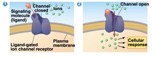
Example: Nicotine and Acetylcholine Receptors
The acetylcholine receptor is a ligand-gated ion channel that can be activated by nicotine, converting chemical signals into electrical signals. 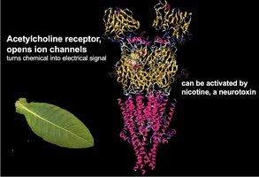
Synaptic Signaling
Synaptic signaling involves the release of neurotransmitters, which bind to ion channel receptors on the postsynaptic cell. 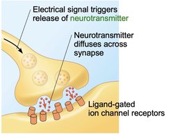
Intracellular Receptors
Intracellular Receptor Function
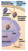
Steroid and thyroid hormones can cross the cell membrane and bind to intracellular receptors. The hormone-receptor complex acts as a transcription factor, regulating gene expression. - Key Point 1: Intracellular receptors are located in the cytoplasm or nucleus. - Key Point 2: Activated complexes directly influence DNA transcription. 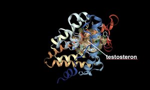
Summary of Receptors
Classification of Cell Signaling Receptors
Receptor Type | Location | Ligand Type | Main Function |
|---|---|---|---|
GPCR | Membrane | Hydrophilic | Signal transduction, amplification |
RTK | Membrane | Hydrophilic | Growth, differentiation |
Ion Channel | Membrane | Hydrophilic | Ion flow, electrical signaling |
Intracellular | Cytoplasm/Nucleus | Hydrophobic | Gene regulation |
Apoptosis (Programmed Cell Death)
Concept and Importance of Apoptosis
Apoptosis is a controlled process by which cells are purposefully eliminated. It is essential for development, maintenance, and defense against damaged or infected cells. - Key Point 1: Apoptosis integrates multiple cell-signaling pathways. - Key Point 2: It prevents uncontrolled cell growth and maintains tissue health. 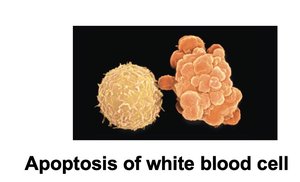
Mechanism of Apoptosis
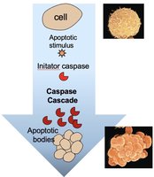
Apoptosis involves the activation of proteases called caspases, which disassemble the cell and package its components into vesicles for digestion by scavenger cells. - Key Point 1: Caspases initiate and execute cell disassembly. - Key Point 2: Apoptotic bodies are formed and removed efficiently. 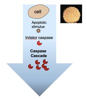
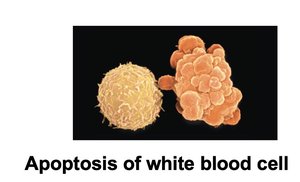
Apoptosis in Development
Apoptosis shapes tissues during development, such as the formation of fingers and toes by removing interdigital cells. 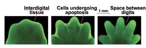
Dysregulation of Apoptosis
Abnormal apoptosis is implicated in diseases such as Parkinson's, Alzheimer's, and cancer. Additional info: Apoptosis is tightly regulated; both excessive and insufficient apoptosis can lead to disease.
