 Indietro
Structure and Function of Prokaryotic Cells
Guida di studio - Note intelligenti
 Appunti personalizzati basati sui tuoi materiali, ampliati con definizioni chiave, esempi e contesto.
Prokaryotic Cell Structure
Overview of Prokaryotic Cells
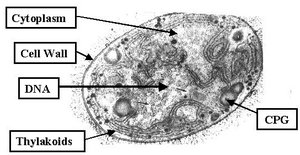
Prokaryotic cells, which include bacteria and archaea, are characterized by their lack of a membrane-bound nucleus and organelles. Despite their simplicity, they possess a variety of specialized structures that enable them to thrive in diverse environments. - Chromosome: Usually a single, circular DNA molecule located in the nucleoid region. - Plasmids: Small, circular DNA molecules that carry additional genes. - Ribosomes: Sites of protein synthesis, composed of RNA and protein. - Cytoplasm: The internal fluid containing all cellular components. 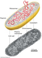
Comparison of Bacterial and Archaeal Phospholipids
The phospholipid composition of prokaryotic membranes differs between bacteria and archaea. - Bacterial phospholipids: Fatty acids bound to glycerol. - Archaeal phospholipids: Branched isoprenoid chains bound to glycerol.
Genetic Material Organization
Nucleoid and DNA Supercoiling
The nucleoid is the region where the chromosome is located. Prokaryotic DNA is often supercoiled to fit within the cell. - Supercoiling: DNA is tightly packed to conserve space and facilitate replication and transcription. - Plasmids: Often supercoiled and provide additional genetic capabilities. 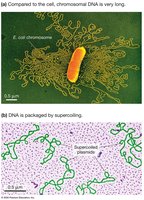
Protein Synthesis
Ribosomes in Prokaryotes
Ribosomes are essential for protein synthesis and are found throughout the cytoplasm. - Structure: Composed of large and small subunits. - Function: Translate mRNA into proteins. - Bacterial vs. Archaeal ribosomes: Similar in size and function, but differ in primary structure.
Cytoskeleton and Cell Shape
Role of the Cytoskeleton
Prokaryotes possess a cytoskeleton made of protein filaments. - Functions: Cell division, maintenance of cell shape, and structural support.
Internal Membrane Complexes
Photosynthetic Membranes
Some prokaryotes, especially photosynthetic species, have internal membrane systems. - Function: Convert sunlight into chemical energy. - Structure: Membranes contain enzymes and pigments necessary for photosynthesis. 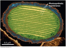
Organelles and Inclusion Bodies
Specialized Internal Compartments
Certain prokaryotes have organelles for specialized functions. - Examples: Storage of calcium ions, magnetite crystals for navigation, and concentration of enzymes.
Inclusion Bodies
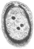
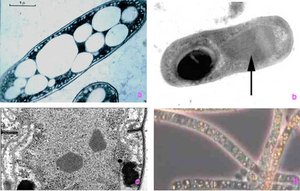
Inclusion bodies are granules of organic or inorganic material used for storage. - Organic inclusion bodies: Carboxysomes (contain Rubisco for photosynthesis), gas vesicles (buoyancy), cyanophycin granules (store nitrogen). - Inorganic inclusion bodies: Store carbon, energy, or other substances. 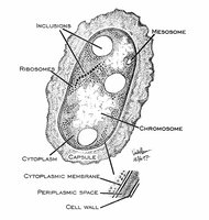
Cell Wall Structure and Function
Composition and Role of the Cell Wall
The cell wall is a protective exoskeleton that maintains cell shape and rigidity. - Peptidoglycan: Main structural component in bacteria, composed of repeating units of N-acetylglucosamine (NAG) and N-acetylmuramic acid (NAM) linked by polypeptides. - Function: Protects against environmental stress and toxic substances, contributes to pathogenicity, and is a target for antibiotics. 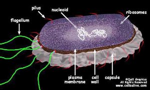
Peptidoglycan Structure
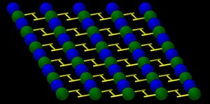
Peptidoglycan forms a mesh-like layer outside the plasma membrane. - Structure: Alternating NAG and NAM sugars form the backbone, cross-linked by peptide chains. - Antibiotics: Some disrupt peptidoglycan cross-linking, causing cell lysis. 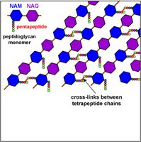
Gram Staining and Cell Wall Differences
Gram-Positive vs. Gram-Negative Bacteria
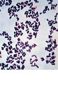
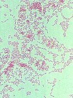
The Gram stain differentiates bacteria based on cell wall structure. - Gram-positive: Thick peptidoglycan layer, stains purple. - Gram-negative: Thin peptidoglycan layer, outer membrane with lipids, stains pink. - Clinical relevance: Gram status affects susceptibility to antibiotics and disease mechanisms. 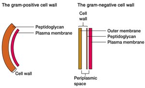
External Structures for Movement and Attachment
Flagella and Fimbriae
Many prokaryotes possess external appendages for movement and attachment. - Flagella: Long, whip-like structures that rotate to propel the cell. - Fimbriae: Short, needle-like projections that facilitate attachment to surfaces or other cells. 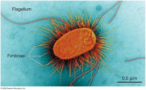
Summary Table: Prokaryotic Cell Structures and Functions
Structure | Function |
|---|---|
Chromosome | Genetic information storage |
Plasmid | Accessory genes, often for antibiotic resistance |
Ribosome | Protein synthesis |
Cytoskeleton | Cell shape, division |
Cell wall | Protection, shape, rigidity |
Flagella | Movement |
Fimbriae | Attachment |
Inclusion bodies | Storage of nutrients |
Photosynthetic membranes | Energy conversion |
Additional info: The notes above expand on the original lecture points with academic context, definitions, and examples to ensure completeness and clarity for General Biology students.
