 Indietro
Study Guide: Archaea and Bacteria Lab (General Biology)
Guida di studio - Note intelligenti
 Appunti personalizzati basati sui tuoi materiali, ampliati con definizioni chiave, esempi e contesto.
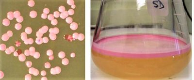
Q1. What is the predominant color of Halobacterium colonies, and what color are the mutant colonies?
Background
Topic: Microbial Colony Morphology and Pigmentation
This question tests your ability to observe and describe the appearance of microbial colonies, specifically Halobacterium, and understand the biological significance of pigment mutations.
Key Terms:
Colony: A visible mass of microorganisms originating from a single cell.
Carotenoid pigments: Molecules that protect DNA from UV radiation.
Mutant: An organism with a genetic change affecting its phenotype.
Step-by-Step Guidance
Examine Figure 1.4, which shows Halobacterium colonies on agar and in broth culture.
Identify the predominant color of the colonies in both the agar and broth images.
Look for colonies that differ in color from the majority—these are the mutants.
Recall that mutant colors are due to carotenoid pigments, which serve a protective function.
Describe the observed colors and consider why these pigments are important for Halobacterium's survival in high-salinity environments.
Try solving on your own before revealing the answer!
Final Answer:
The predominant color of Halobacterium colonies is pink. The mutant colonies are orange or red. These colors are due to carotenoid pigments, which protect the organism's DNA from harmful UV radiation in their extreme environments.
Q2. What type of archaean is Halobacterium, and what special requirements would be needed to culture it?
Background
Topic: Microbial Classification and Environmental Adaptations
This question tests your understanding of extremophiles and their unique environmental needs, specifically Halobacterium as a halophile.
Key Terms:
Halophile: An organism that thrives in high-salt environments.
Culture: The process of growing microorganisms under controlled conditions.
Step-by-Step Guidance
Recall the definition of halophiles and their environmental requirements.
Review the information about Halobacterium's need for high salinity (above 9%).
Consider what would be necessary in a laboratory setting to successfully culture Halobacterium.
Think about the comparison to mammalian cell saline content (0.9%) and why Halobacterium needs much higher salt concentrations.
List the special requirements for culturing halophiles, such as the composition of the growth medium.
Try solving on your own before revealing the answer!
Final Answer:
Halobacterium is a halophile, meaning it requires high salt concentrations to survive. To culture it, you need a growth medium with salinity above 9%, much higher than typical media used for other organisms. This ensures the organism can thrive and prevents contamination by non-halophilic microbes.
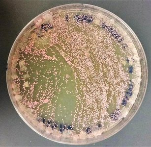
Q3. Do you see evidence of the special requirements on the agar in the Petri dish? What do you see around the edges?
Background
Topic: Microbial Growth and Environmental Adaptations
This question asks you to observe the physical evidence of Halobacterium's special requirements on a Petri dish, specifically signs of high salinity.
Key Terms:
Agar: A gel-like substance used as a growth medium for microorganisms.
Petri dish: A shallow dish used to culture microorganisms.
Salinity: The concentration of salt in a solution.
Step-by-Step Guidance
Examine the Petri dish image (Figure 1.5) for any visible signs of salt or other unusual features.
Look closely at the edges of the agar for crystalline deposits or changes in texture.
Consider how high salinity might manifest visually in the growth medium.
Think about why these features are important for Halobacterium's survival.
Try solving on your own before revealing the answer!
Final Answer:
Yes, evidence of special requirements is visible on the agar. Around the edges, you can see crystalline salt deposits, which indicate the high salinity needed for Halobacterium to grow. These deposits are a direct result of the organism's need for a salty environment.
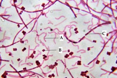
Q4. Identify the shapes labeled A, B, and C in the mixed bacteria image.
Background
Topic: Bacterial Morphology
This question tests your ability to recognize and distinguish between the three basic shapes of bacteria: coccus, bacillus, and spirillum.
Key Terms:
Coccus: Spherical-shaped bacteria.
Bacillus: Rod-shaped bacteria.
Spirillum: Spiral-shaped bacteria.
Step-by-Step Guidance
Examine the mixed bacteria image for three distinct shapes.
Identify which labeled group (A, B, C) corresponds to each shape: spherical, rod, or spiral.
Recall the definitions of coccus, bacillus, and spirillum.
Match the labels to the correct bacterial shape based on their appearance.
Try solving on your own before revealing the answer!
Final Answer:
A: Spirillum (spiral-shaped), B: Bacillus (rod-shaped), C: Coccus (spherical-shaped). These are the three basic shapes of bacteria commonly observed under the microscope.
Q5. How would you expect the following genera of bacteria to appear based on their names: Staphylococcus, Streptobacillus, Diplococcus, Streptococcus?
Background
Topic: Bacterial Nomenclature and Cell Arrangement
This question tests your understanding of how bacterial genus names reflect cell shape and arrangement.
Key Terms:
Staphylo-: Cluster arrangement
Strepto-: Chain arrangement
Diplo-: Pair arrangement
Coccus: Spherical shape
Bacillus: Rod shape
Step-by-Step Guidance
Break down each genus name into its prefix and root.
Recall what each prefix indicates about cell arrangement.
Combine the prefix meaning with the root shape to predict the appearance.
Visualize or sketch how each genus would look under the microscope.
Try solving on your own before revealing the answer!
Final Answer:
Staphylococcus: Spherical cells in clusters.
Streptobacillus: Rod-shaped cells in chains.
Diplococcus: Spherical cells in pairs.
Streptococcus: Spherical cells in chains.
The prefixes describe the arrangement, and the root describes the shape.
Q6. Complete Table 1 for the five Gram-stained bacterial species: Klebsiella pneumoniae, Neisseria gonorrhoeae, Pseudomonas aeruginosa, Bacillus anthracis, Staphylococcus epidermidis.
Background
Topic: Bacterial Classification, Gram Staining, and Human Impact
This question tests your ability to identify bacterial shape, cell arrangement, Gram reaction, and their significance to human health.
Key Terms:
Gram stain: A method to differentiate bacteria based on cell wall structure.
Gram-positive: Stains purple, thick peptidoglycan wall.
Gram-negative: Stains pink, thin peptidoglycan wall.
Pathogen: An organism that causes disease.
Step-by-Step Guidance
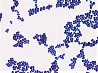
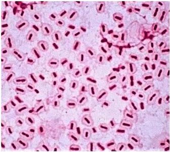
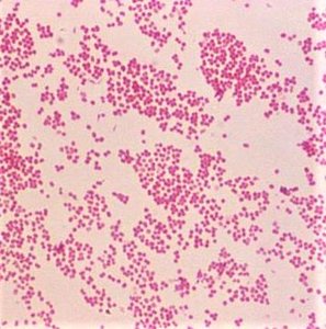
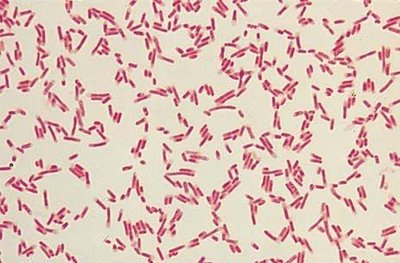
Review the images of each species (Figures 1.5, 1.6, 1.7) for cell shape and arrangement.
Determine the Gram reaction (positive or negative) based on color in the images.
Research or recall the human impact of each species (disease or beneficial effect).
Fill in the table with shape, arrangement, Gram reaction, and human impact for each species.

Try solving on your own before revealing the answer!
Final Answer:
Klebsiella pneumoniae: Shape - bacillus (rod), Organization - single or pairs, Gram-negative, Human Impact - causes pneumonia.
Neisseria gonorrhoeae: Shape - coccus (spherical), Organization - diplococcus (pairs), Gram-negative, Human Impact - causes gonorrhea.
Pseudomonas aeruginosa: Shape - bacillus (rod), Organization - single, Gram-negative, Human Impact - opportunistic infections, especially in immunocompromised patients.
Bacillus anthracis: Shape - bacillus (rod), Organization - chains, Gram-positive, Human Impact - causes anthrax.
Staphylococcus epidermidis: Shape - coccus (spherical), Organization - clusters, Gram-positive, Human Impact - skin infections, usually non-pathogenic but can cause infections in immunocompromised individuals.
Q7. Identify the cell labeled “A” and the cells labeled “B” in Figure 1.9 (Anabaena).
Background
Topic: Cyanobacteria Cell Types and Nitrogen Fixation
This question tests your ability to distinguish between photosynthetic cells and heterocysts in Anabaena, a cyanobacterium.
Key Terms:
Heterocyst: Specialized cell for nitrogen fixation.
Photosynthetic cell: Cell that performs photosynthesis.
Step-by-Step Guidance
Examine Figure 1.9 for two distinct cell types: smaller roundish cells and larger oval cells.
Recall that heterocysts are larger and specialized for nitrogen fixation.
Photosynthetic cells are smaller and more numerous.
Match the labels A and B to the correct cell types based on their appearance and function.
Try solving on your own before revealing the answer!
Final Answer:
A: Heterocyst (nitrogen-fixing cell), B: Photosynthetic cells. Heterocysts are larger and less frequent, while photosynthetic cells are smaller and form the main chain.
Q8. Write the name of each organism studied during this lab under the correct classification: Halophiles, Thermophiles, Methanogens, Gram Negative bacteria, Cyanobacteria, Gram Positive bacteria.
Background
Topic: Prokaryote Classification
This question tests your ability to classify organisms based on their environmental adaptations and Gram reaction.
Key Terms:
Halophile: High salt requirement
Thermophile: High temperature requirement
Methanogen: Produces methane
Gram-negative: Pink stain
Gram-positive: Purple stain
Cyanobacteria: Photosynthetic bacteria
Step-by-Step Guidance
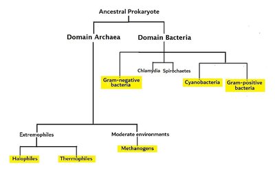
Review the organisms studied: Halobacterium, Pyrococcus, Methanococcus, Klebsiella pneumoniae, Neisseria gonorrhoeae, Pseudomonas aeruginosa, Bacillus anthracis, Staphylococcus epidermidis, Oscillatoria, Anabaena.
Assign each organism to the correct classification based on their characteristics.
Use the phylogenetic tree and information from the lab to help with classification.
Try solving on your own before revealing the answer!
Final Answer:
Halophiles: Halobacterium
Thermophiles: Pyrococcus
Methanogens: Methanococcus
Gram Negative bacteria: Klebsiella pneumoniae, Neisseria gonorrhoeae, Pseudomonas aeruginosa
Cyanobacteria: Oscillatoria, Anabaena
Gram Positive bacteria: Bacillus anthracis, Staphylococcus epidermidis
Q9. State a hypothesis and null hypothesis for the experiment testing the effectiveness of disinfecting agents on bacterial growth.
Background
Topic: Scientific Method and Experimental Design
This question tests your ability to formulate hypotheses for an experiment comparing antibacterial soap and hand sanitizer.
Key Terms:
Hypothesis: An educated guess about the outcome.
Null hypothesis: A statement that there will be no effect or difference.
Step-by-Step Guidance
Consider the purpose of the experiment: comparing the effectiveness of two disinfecting agents.
Formulate a hypothesis predicting which agent will be more effective, or if both will be equally effective.
Write a null hypothesis stating that there will be no difference in effectiveness.
Try solving on your own before revealing the answer!
Final Answer:
Hypothesis: Hand sanitizer containing 70% isopropyl alcohol will be more effective at reducing bacterial growth than antibacterial soap.
Null Hypothesis: There will be no difference in bacterial growth reduction between hand sanitizer and antibacterial soap.
