 Indietro
Study Guide: Neurons, Synapses, Signaling, and Nervous/Sensory Systems
Guida di studio - Note intelligenti
 Appunti personalizzati basati sui tuoi materiali, ampliati con definizioni chiave, esempi e contesto.
Q1. What is a neuron?
Background
Topic: Structure and Function of Neurons
This question is testing your understanding of the basic unit of the nervous system and its role in transmitting information.
Key Terms:
Neuron: A specialized cell that transmits nerve impulses.
Impulse: An electrical signal that travels along the neuron.
Step-by-Step Guidance
Recall that neurons are the fundamental cells of the nervous system.
Think about the main function of neurons: transmitting electrical and chemical signals.
Consider the structural features of a neuron, such as dendrites, axon, and cell body.
Try solving on your own before revealing the answer!
Final Answer:
A neuron is a specialized cell in the nervous system that transmits electrical and chemical signals to other cells. It consists of a cell body, dendrites, and an axon.
Q2. Neurons can be placed into three groups, based on their location and function. Fill in the functions for Sensory Neurons, Interneurons, and Motor Neurons.
Background
Topic: Types of Neurons
This question is testing your ability to distinguish between the three main types of neurons and their roles in the nervous system.
Key Terms:
Sensory Neurons: Carry information from sensory receptors to the CNS.
Interneurons: Process information within the CNS.
Motor Neurons: Carry instructions from the CNS to effectors (muscles/glands).
Step-by-Step Guidance
Recall the pathway of information in the nervous system: from sensory input to processing to response.
Think about which neurons are responsible for detecting stimuli (sensory), which process information (interneurons), and which cause a response (motor).
Match each type of neuron to its function based on its location and role.
Try solving on your own before revealing the answer!
Final Answer:
Sensory Neurons: Carry information from sensory receptors to the central nervous system.
Interneurons: Integrate and process information within the central nervous system.
Motor Neurons: Transmit signals from the central nervous system to muscles or glands to elicit a response.
Q3. Which division of the nervous system includes the brain and spinal cord?
Background
Topic: Organization of the Nervous System
This question is testing your knowledge of the structural divisions of the nervous system.
Key Terms:
Central Nervous System (CNS): Includes the brain and spinal cord.
Peripheral Nervous System (PNS): Includes nerves outside the CNS.
Step-by-Step Guidance
Recall the two main divisions of the nervous system: CNS and PNS.
Identify which division contains the brain and spinal cord.
Try solving on your own before revealing the answer!
Final Answer:
The central nervous system (CNS) includes the brain and spinal cord.
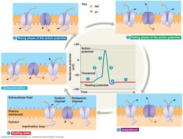
Q4. Label the following elements of this figure: cell body, dendrites, axon, synapse, presynaptic cell, postsynaptic cell, synaptic vesicles, synaptic terminal, and neurotransmitter.
Background
Topic: Structure of Neurons and Synapses
This question is testing your ability to identify and label the key parts of a neuron and its synaptic connections.
Key Terms:
Cell Body: Contains the nucleus and organelles.
Dendrites: Receive signals from other neurons.
Axon: Transmits signals away from the cell body.
Synapse: Junction between two neurons.
Presynaptic Cell: The neuron sending the signal.
Postsynaptic Cell: The neuron receiving the signal.
Synaptic Vesicles: Contain neurotransmitters.
Synaptic Terminal: End of the axon where neurotransmitters are released.
Neurotransmitter: Chemical messenger released at the synapse.
Step-by-Step Guidance
Examine the diagram and locate the cell body, dendrites, and axon.
Identify the synapse, which is the gap between two neurons.
Label the presynaptic and postsynaptic cells based on the direction of signal transmission.
Find the synaptic terminal and synaptic vesicles at the end of the axon.
Locate where neurotransmitters are released into the synaptic cleft.
Try solving on your own before revealing the answer!
Final Answer:
Cell body: Central part of the neuron containing the nucleus.
Dendrites: Branch-like structures extending from the cell body.
Axon: Long extension leading away from the cell body.
Synapse: Gap between the axon terminal and the next cell.
Presynaptic cell: The neuron sending the signal.
Postsynaptic cell: The neuron receiving the signal.
Synaptic vesicles: Small sacs in the synaptic terminal containing neurotransmitters.
Synaptic terminal: End of the axon where neurotransmitters are released.
Neurotransmitter: Chemical messenger released into the synapse.
Q5. What is shown in the previous figure?
Background
Topic: Neuron Structure and Synaptic Transmission
This question is testing your ability to interpret a diagram of neuron anatomy and synaptic function.
Key Terms:
Neuron: Nerve cell.
Synapse: Junction between neurons.
Step-by-Step Guidance
Review the labeled parts of the neuron in the diagram.
Consider the overall function of the structures shown.
Think about how the diagram illustrates the process of signal transmission between neurons.
Try solving on your own before revealing the answer!
Final Answer:
The figure shows the structure of two neurons and their synaptic connection, illustrating how signals are transmitted from one neuron to another.
Q6. What is indicated by the red arrows in the main part of the figure you labeled?
Background
Topic: Direction of Signal Transmission
This question is testing your understanding of how electrical impulses travel through a neuron.
Key Terms:
Action Potential: Electrical signal moving along the axon.
Synaptic Transmission: Movement of signal from one neuron to another.
Step-by-Step Guidance
Look at the direction of the arrows in the diagram.
Recall that action potentials travel from the cell body down the axon to the synaptic terminal.
Consider what happens at the synapse when the signal reaches the terminal.
Try solving on your own before revealing the answer!
Final Answer:
The red arrows indicate the direction of the action potential, showing how the electrical signal travels from the cell body along the axon to the synaptic terminal.
Q7. What are glial cells?
Background
Topic: Supporting Cells in the Nervous System
This question is testing your knowledge of the types and functions of glial cells.
Key Terms:
Glial Cells: Non-neuronal cells that support and protect neurons.
Myelin Sheath: Insulating layer formed by some glial cells.
Step-by-Step Guidance
Recall that glial cells are not neurons but play essential roles in the nervous system.
Think about the functions of glial cells, such as providing support, insulation, and nutrients to neurons.
Consider examples of glial cells, like Schwann cells and oligodendrocytes.
Try solving on your own before revealing the answer!
Final Answer:
Glial cells are supporting cells in the nervous system that provide structural support, insulation, and nutrients to neurons. They also help maintain homeostasis and form myelin sheaths.
