 Indietro
The Cell Cycle and Mitosis: Structure, Function, and Regulation
Guida di studio - Note intelligenti
 Appunti personalizzati basati sui tuoi materiali, ampliati con definizioni chiave, esempi e contesto.
The Cell Cycle
Overview of the Cell Cycle
The cell cycle is the series of events that cells undergo as they grow and divide. It consists of interphase (where the cell grows and DNA is replicated) and the mitotic (M) phase (where cell division occurs). - Interphase includes three subphases: G1 (cell growth), S (DNA synthesis), and G2 (preparation for division). - Mitotic Phase includes mitosis (division of the nucleus) and cytokinesis (division of the cytoplasm). - Cells that cease division enter a non-dividing state called G0. 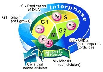
Functions of Cell Division
Cell division is essential for reproduction, growth, development, and tissue renewal. - Asexual reproduction in unicellular organisms produces identical offspring. - Growth and development in multicellular organisms occurs through repeated cell divisions. - Tissue renewal replaces damaged or dead cells. 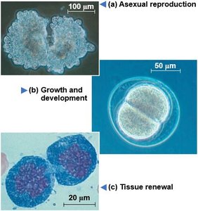
Genetic Material and Chromosomes
Organization of Genetic Material
The genetic material of a cell is organized into chromosomes, which are composed of DNA and associated proteins (chromatin). - Genome: All DNA in a cell. - Chromatin: Complex of DNA and protein; condenses to form chromosomes during cell division. - Somatic cells are diploid (2n), containing two sets of chromosomes. - Gametes are haploid (n), containing one set of chromosomes. 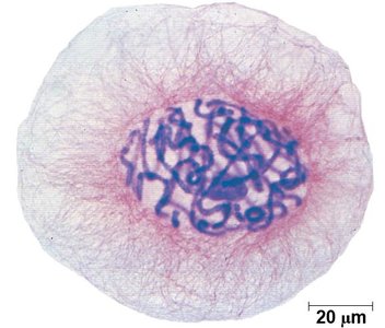
Chromosome Duplication and Distribution
Before cell division, chromosomes are duplicated, forming two sister chromatids joined at the centromere. - Sister chromatids are identical copies of a chromosome. - Centromere is the region where chromatids are joined. - During mitosis, sister chromatids separate, becoming daughter chromosomes. 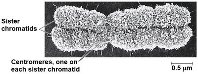
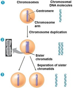
Phases of the Cell Cycle
Interphase
Interphase is the longest phase of the cell cycle, consisting of G1, S, and G2. - G1 phase: Cell grows and synthesizes proteins. - S phase: DNA is replicated. - G2 phase: Cell prepares for division by synthesizing proteins and organelles. 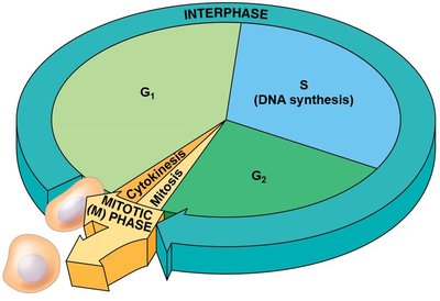
Mitosis: Stages and Events
Mitosis is conventionally divided into five stages: prophase, prometaphase, metaphase, anaphase, and telophase.
Prophase and Prometaphase
- Chromatin condenses into visible chromosomes. - Mitotic spindle forms as centrosomes move to opposite poles. - Nuclear envelope breaks down; spindle fibers attach to kinetochores. 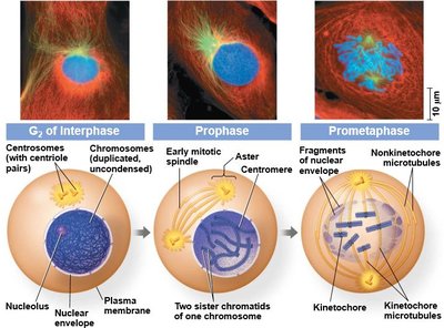
Metaphase, Anaphase, Telophase, and Cytokinesis
- Metaphase: Chromosomes align at the metaphase plate. - Anaphase: Sister chromatids are pulled apart to opposite poles. - Telophase: Chromosomes decondense; nuclear envelope reforms. - Cytokinesis: Cytoplasm divides, forming two daughter cells. 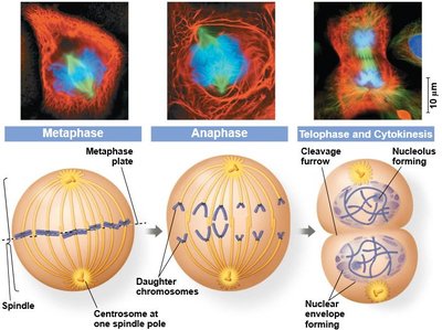
The Mitotic Spindle
Structure and Function
The mitotic spindle is a structure made of microtubules that orchestrates chromosome movement during mitosis. - Includes centrosomes, spindle microtubules, and asters. - Microtubules attach to kinetochores on chromosomes. 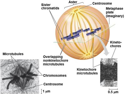
Mechanism of Chromosome Movement
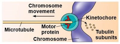
- During anaphase, cohesins are cleaved, and sister chromatids move toward opposite poles. - Motor proteins at kinetochores "walk" chromosomes along microtubules. - Microtubules depolymerize at kinetochore ends, facilitating movement. 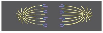
Cytokinesis
Animal vs. Plant Cells
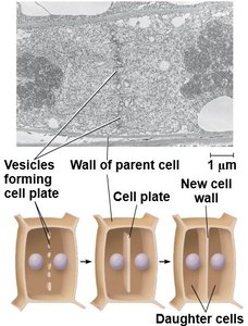
Cytokinesis differs between animal and plant cells. - Animal cells: Form a cleavage furrow, pinching the cell into two. - Plant cells: Form a cell plate, which develops into a new cell wall. 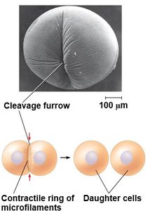
Binary Fission in Bacteria
Mechanism of Binary Fission
Prokaryotes reproduce by binary fission, a simpler process than mitosis. - Chromosome replicates, and daughter chromosomes move apart. - Plasma membrane pinches inward, dividing the cell.
Regulation of the Cell Cycle
Cell Cycle Control System
The cell cycle is regulated by a molecular control system, including checkpoints at G1, G2, and M phases. - Cyclins and cyclin-dependent kinases (Cdks) are key regulatory proteins. - MPF (maturation-promoting factor) is a cyclin-Cdk complex that triggers entry into mitosis.
Checkpoints and Signals
- Internal signals ensure proper chromosome attachment before anaphase. - External signals include growth factors, density-dependent inhibition, and anchorage dependence.
Cancer and Loss of Cell Cycle Control
Cancer Cells
Cancer cells evade normal cell cycle controls, leading to uncontrolled division. - May produce their own growth factors or have abnormal control systems. - Can form tumors, which may be benign or malignant (capable of metastasis).
Cancer Treatment
- Treatments include radiation, chemotherapy (e.g., Taxol), and surgery. - Advances in cell cycle understanding have led to more personalized therapies. Additional info: The notes expand on the original content by providing definitions, mechanisms, and context for each process, ensuring completeness and academic quality for exam preparation.
