 Indietro
The Cell Cycle and Molecular Biology of Cancer: Study Notes for General Biology
Guida di studio - Note intelligenti
 Appunti personalizzati basati sui tuoi materiali, ampliati con definizioni chiave, esempi e contesto.
Chapter 12: The Cell Cycle
Big Idea and Overview
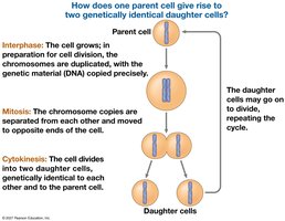
The cell cycle is fundamental to understanding how organisms grow, reproduce, and maintain their tissues. Cell division is the process by which a single cell gives rise to two genetically identical daughter cells, ensuring continuity of life and accurate transmission of genetic material.
Key Roles of Cell Division
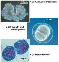
Asexual reproduction: Single-celled organisms reproduce by cell division.
Growth and development: Multicellular organisms develop from a single cell through repeated cell divisions.
Tissue renewal: Cell division repairs and renews tissues in multicellular organisms.
Concept 12.1: Genetically Identical Daughter Cells
Cellular Organization of Genetic Material
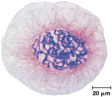
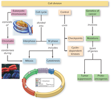
All the DNA in a cell constitutes its genome. Prokaryotes typically have a single DNA molecule, while eukaryotes have multiple DNA molecules organized into chromosomes. Chromosomes are composed of chromatin, a complex of DNA and protein that condenses during cell division.
Somatic cells: Non-reproductive cells with two sets of chromosomes.
Gametes: Reproductive cells (sperm and eggs) with half the chromosome number of somatic cells.
Chromosome Structure and Replication
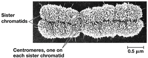
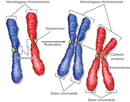
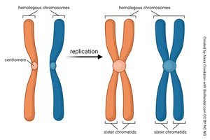
Before cell division, chromosomes are duplicated, resulting in two sister chromatids joined at a centromere. During division, sister chromatids separate, becoming individual chromosomes.
Concept 12.2: The Cell Cycle Phases
Phases of the Cell Cycle
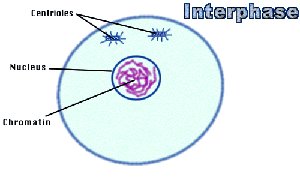
The cell cycle consists of interphase (cell growth and DNA replication) and the mitotic (M) phase (mitosis and cytokinesis).
Interphase: Divided into G1 (growth), S (DNA synthesis), and G2 (preparation for division).
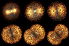
Mitosis: Division of the nucleus, conventionally broken into five stages: prophase, prometaphase, metaphase, anaphase, and telophase.
Cytokinesis: Division of the cytoplasm, resulting in two daughter cells.
Stages of Mitosis
Prophase: Chromosomes condense, spindle apparatus forms, nuclear envelope breaks down.
Prometaphase: Microtubules attach to kinetochores, chromosomes begin moving to the cell center.
Metaphase: Chromosomes align at the metaphase plate.
Anaphase: Sister chromatids separate and move to opposite poles.
Telophase: Nuclear envelope reforms, chromosomes decondense.
Concept 12.3: Regulation of the Cell Cycle
Cell Cycle Control System
The cell cycle is regulated by checkpoints, cyclins, cyclin-dependent kinases (Cdks), and maturation-promoting factor (MPF). Checkpoints ensure proper progression and prevent errors.
G1 checkpoint: Determines if the cell will proceed to division or enter G0 (non-dividing state).
G2 checkpoint: Ensures DNA replication is complete and undamaged.
M checkpoint: Ensures all chromosomes are properly attached to the spindle before anaphase.
Concept 18.5: Molecular Biology of Cancer
Genetic Changes Leading to Cancer
Cancer results from mutations that disrupt normal cell cycle control. Key genes involved include proto-oncogenes (which can become oncogenes) and tumor-suppressor genes.
Proto-oncogenes: Normally stimulate cell growth; mutations can convert them to oncogenes, leading to uncontrolled division.
Tumor-suppressor genes: Normally inhibit cell division; mutations can reduce their function, contributing to cancer.
Ras proto-oncogene: Mutations can cause hyperactive Ras protein, stimulating excessive cell division.
p53 tumor suppressor gene: Mutations prevent suppression of the cell cycle, allowing cells with DNA damage to divide.
Multistep Model of Cancer Development
Multiple mutations are typically required for cancer to develop, often involving both activation of oncogenes and loss of tumor-suppressor gene function. Inherited mutations (such as in BRCA1 or BRCA2) can predispose individuals to certain cancers.
Summary Table: Cell Cycle and Cancer Genes
Gene Type | Normal Function | Effect of Mutation |
|---|---|---|
Proto-oncogene | Stimulates cell growth | Becomes oncogene, causes uncontrolled division |
Tumor-suppressor gene | Inhibits cell division, repairs DNA | Loss of function, allows excessive division |
Ras | Signal transduction for growth | Hyperactive protein, increased division |
p53 | DNA repair, cell cycle arrest, apoptosis | Loss of suppression, cells with damage divide |
Conclusion
Understanding the cell cycle and its regulation is essential for grasping how organisms grow, reproduce, and maintain their tissues. Disruptions in cell cycle control can lead to cancer, highlighting the importance of molecular checkpoints and genetic regulation.
