 Indietro
The Cell Cycle: Mechanisms and Regulation
Guida di studio - Note intelligenti
 Appunti personalizzati basati sui tuoi materiali, ampliati con definizioni chiave, esempi e contesto.
The Cell Cycle
Key Roles of Cell Division
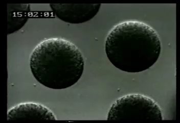
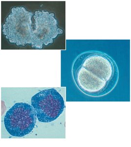
Cell division is fundamental to the continuity of life, enabling organisms to reproduce, grow, renew, and repair tissues. In unicellular organisms, division of one cell reproduces the entire organism, while in multicellular eukaryotes, cell division supports development, growth, and tissue repair.
Reproduction: Single-celled organisms reproduce by cell division.
Growth and Development: Multicellular organisms grow and develop from a single fertilized cell through repeated cell divisions.
Tissue Renewal: Cell division replaces cells that are lost due to normal wear and tear or injury.
Genetic Material Organization
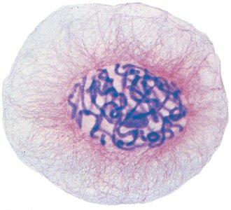
All the DNA in a cell constitutes its genome. In eukaryotes, DNA is packaged into multiple chromosomes, each consisting of chromatin—a complex of DNA and protein. Somatic cells (nonreproductive) have two sets of chromosomes, while gametes (sperm and eggs) have half as many.
Chromatin: The material of which eukaryotic chromosomes are composed; consists of DNA and proteins.
Chromosome: A structure carrying genetic material, found in the nucleus of eukaryotic cells.
Genome: The complete set of genes or genetic material present in a cell or organism.
Chromosome Duplication and Distribution
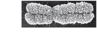
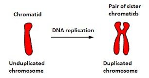
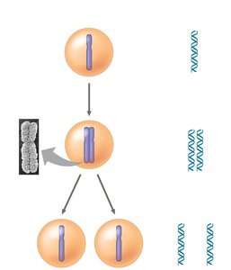
Before cell division, chromosomes are duplicated. Each duplicated chromosome consists of two sister chromatids joined at a region called the centromere. During division, sister chromatids separate and are distributed to daughter cells.
Sister chromatids: Identical copies of a chromosome connected by a centromere.
Centromere: The region where sister chromatids are most closely attached.
Phases of the Cell Cycle
Overview of the Cell Cycle
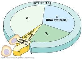
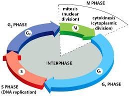
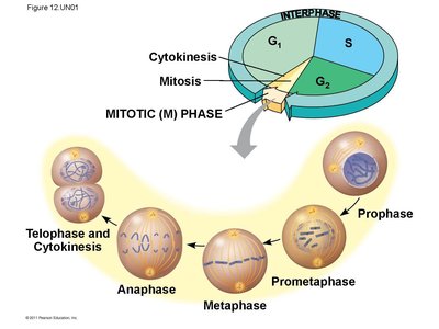
The cell cycle consists of two main phases: Interphase and the Mitotic (M) phase. Interphase is subdivided into G1 (first gap), S (synthesis), and G2 (second gap) phases. The M phase includes mitosis (nuclear division) and cytokinesis (cytoplasmic division).
G1 phase: Cell grows and carries out normal functions.
S phase: DNA is replicated.
G2 phase: Cell prepares for division.
M phase: Mitosis and cytokinesis occur.
Mitosis: Phases and Events
Mitosis is divided into several phases: prophase, prometaphase, metaphase, anaphase, and telophase. Each phase is characterized by specific events that ensure accurate chromosome segregation.
Prophase: Chromosomes condense, spindle apparatus begins to form.
Prometaphase: Nuclear envelope fragments, spindle microtubules attach to kinetochores.
Metaphase: Chromosomes align at the metaphase plate.
Anaphase: Sister chromatids separate and move toward opposite poles.
Telophase: Nuclear envelopes reform around chromosomes at each pole.
Cytokinesis: Division of the cytoplasm, forming two daughter cells.
The Mitotic Spindle
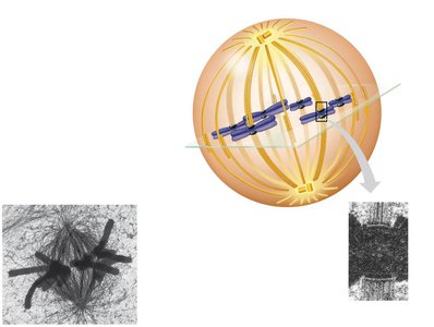
The mitotic spindle is a structure made of microtubules that orchestrates the movement of chromosomes during mitosis. In animal cells, spindle formation begins at the centrosomes, which replicate and migrate to opposite poles. The spindle includes centrosomes, spindle microtubules, and asters.
Centrosome: Microtubule organizing center in animal cells.
Kinetochore: Protein complex at the centromere where spindle fibers attach.
Aster: Radial array of short microtubules extending from each centrosome.
Cytokinesis
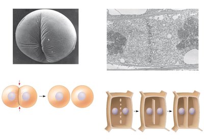
Cytokinesis divides the cytoplasm, resulting in two daughter cells. In animal cells, this occurs by cleavage, forming a cleavage furrow. In plant cells, a cell plate forms, which develops into a new cell wall.
Cleavage furrow: Indentation that begins the process of cleavage in animal cells.
Cell plate: Structure that forms in plant cells during cytokinesis, leading to the formation of a new cell wall.
Regulation of the Cell Cycle
Cell Cycle Control System
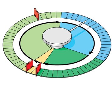
The cell cycle is regulated by a molecular control system with checkpoints at G1, G2, and M phases. These checkpoints ensure that critical processes are completed before the cell proceeds to the next phase. The system is regulated by internal and external signals.
Checkpoint: Control point where stop and go-ahead signals regulate the cycle.
G0 phase: Nondividing state that a cell may enter if it does not receive a go-ahead signal at the G1 checkpoint.
Cyclins, Cdks, and MPF
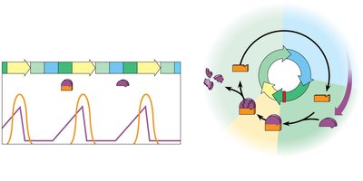
Two types of regulatory proteins, cyclins and cyclin-dependent kinases (Cdks), control the cell cycle. The activity of Cdks fluctuates with the concentration of cyclins. The maturation-promoting factor (MPF) is a cyclin-Cdk complex that triggers passage through the G2 checkpoint into M phase.
Cyclin: Protein whose concentration cycles throughout the cell cycle.
Cdk: Enzyme that, when combined with cyclin, can phosphorylate target proteins to regulate the cell cycle.
MPF: Cyclin-Cdk complex that promotes mitosis.
Internal and External Signals
Internal signals, such as proper chromosome attachment to the spindle, and external signals, such as growth factors, regulate cell division. Density-dependent inhibition and anchorage dependence are mechanisms that prevent uncontrolled cell growth.
Growth factor: Protein that stimulates cell division.
Density-dependent inhibition: Cells stop dividing when crowded.
Anchorage dependence: Cells must be attached to a substratum to divide.
Loss of Cell Cycle Controls in Cancer Cells
Cancer cells do not respond to normal cell cycle controls. They may divide uncontrollably, form tumors, and invade other tissues (metastasis). Cancer treatments often target the cell cycle to inhibit tumor growth.
Transformation: Process by which a normal cell becomes cancerous.
Benign tumor: Abnormal cells remain at the original site.
Malignant tumor: Abnormal cells invade surrounding tissues and can metastasize.
Binary Fission in Bacteria
Mechanism of Binary Fission
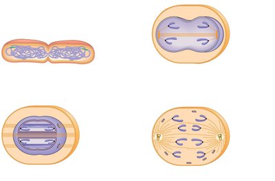
Prokaryotes reproduce by binary fission, a process in which the chromosome replicates and the cell divides into two genetically identical daughter cells. This process is simpler than mitosis and is thought to be the evolutionary precursor to eukaryotic cell division.
Origin of replication: Specific site where DNA replication begins in prokaryotes.
Evolution of Mitosis
Mitosis likely evolved from binary fission. Some protists exhibit cell division processes that are intermediate between binary fission and mitosis, supporting this evolutionary relationship.
Additional info: Understanding the cell cycle and its regulation is crucial for fields such as developmental biology, cancer research, and regenerative medicine.
