 Indietro
The Cell Cycle: Structure, Function, and Regulation
Guida di studio - Note intelligenti
 Appunti personalizzati basati sui tuoi materiali, ampliati con definizioni chiave, esempi e contesto.
The Cell Cycle
Overview and Importance of Cell Division
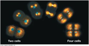
The cell cycle is a fundamental process in biology, responsible for the growth, development, and maintenance of all living organisms. It ensures the continuity of life by enabling cells to reproduce, thereby passing genetic material from one generation to the next.
Cell Division is essential for reproduction in single-celled organisms, embryonic development in multicellular organisms, and tissue renewal and repair in adults.
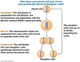
Cell division is highly accurate, ensuring that daughter cells receive identical genetic material.
Organisms distinguish themselves from nonliving matter by their ability to produce more of their own kind through cell division.
Key Roles of Cell Division
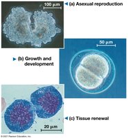
Asexual reproduction: Single-celled organisms reproduce by dividing into new organisms.
Growth and development: Multicellular organisms grow and develop through repeated cell divisions.
Tissue renewal: Fully grown multicellular organisms use cell division to replace damaged or lost cells.
Cellular Organization of Genetic Material
Chromosomes and Chromatin
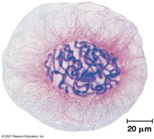
Genetic material in cells is organized into chromosomes, which are composed of DNA and associated proteins. The DNA molecule of a chromosome carries hundreds to thousands of genes.
Genome: All the DNA in a cell; can be a single DNA molecule (prokaryotes) or multiple DNA molecules (eukaryotes).
Chromatin: The complex of DNA and protein that condenses to form chromosomes during cell division.
Somatic cells: Nonreproductive cells with two sets of chromosomes.
Gametes: Reproductive cells (sperm and eggs) with half the chromosome number of somatic cells.
Chromosome Duplication and Distribution
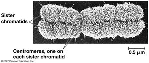
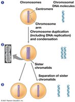
Before cell division, chromosomes are duplicated, resulting in two sister chromatids joined at the centromere. During division, chromatids separate and are distributed to daughter cells.
Sister chromatids: Joined copies of the original chromosome.
Centromere: The region where chromatids are most closely attached.
Phases of the Cell Cycle
Interphase and Mitotic (M) Phase
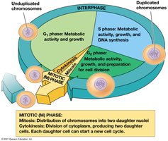
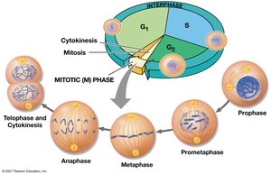
The cell cycle consists of two main phases: interphase and the mitotic (M) phase. Interphase is subdivided into G1, S, and G2 phases, during which the cell grows and DNA is replicated. The M phase includes mitosis and cytokinesis.
G1 phase: Metabolic activity and growth.
S phase: DNA synthesis and chromosome duplication.
G2 phase: Preparation for cell division.
Mitosis: Distribution of chromosomes into two daughter nuclei.
Cytokinesis: Division of cytoplasm, producing two daughter cells.
Stages of Mitosis
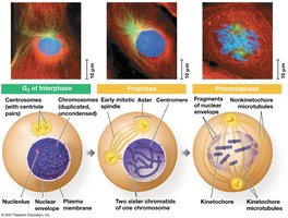
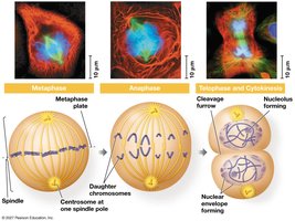
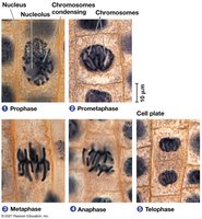
Mitosis is conventionally divided into five stages: prophase, prometaphase, metaphase, anaphase, and telophase. Each stage is characterized by specific events involving chromosome movement and nuclear envelope changes.
Prophase: Chromosomes condense, spindle forms.
Prometaphase: Nuclear envelope fragments, spindle microtubules attach to kinetochores.
Metaphase: Chromosomes align at the metaphase plate.
Anaphase: Sister chromatids separate and move to opposite poles.
Telophase: Nuclear envelope reforms, chromosomes decondense.
The Mitotic Spindle
Structure and Function
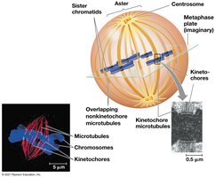
The mitotic spindle is a structure made of microtubules that controls chromosome movement during mitosis. It includes centrosomes, spindle microtubules, and asters.
Centrosome: Microtubule-organizing center that replicates and migrates to opposite ends of the cell.
Aster: Radial array of short microtubules extending from each centrosome.
Kinetochore: Protein complex at the centromere where spindle microtubules attach.
Metaphase plate: Imaginary plane where chromosomes align during metaphase.
Chromosome Movement During Anaphase
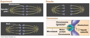
During anaphase, sister chromatids are separated by the action of motor proteins and microtubule depolymerization. Chromosomes are moved toward opposite poles by two mechanisms: the "Pac-man" mechanism and reeling in by motor proteins at the spindle poles.
Separase: Enzyme that cleaves cohesins, allowing chromatids to separate.
Microtubule depolymerization: Shortening of microtubules at kinetochore ends.
Cytokinesis
Mechanisms in Animal and Plant Cells
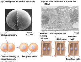
Cytokinesis is the process by which the cytoplasm divides, resulting in two daughter cells. In animal cells, this occurs by cleavage, forming a cleavage furrow. In plant cells, a cell plate forms, eventually becoming the new cell wall.
Cleavage furrow: Shallow groove in the cell surface indicating the start of cytokinesis in animal cells.
Cell plate: Structure formed during cytokinesis in plant cells.
Binary Fission in Bacteria
Prokaryotic Cell Division
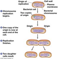
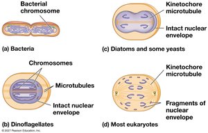
Prokaryotes reproduce by binary fission, a process in which the chromosome replicates and the cell divides into two. This mechanism is simpler than mitosis and is believed to be the evolutionary precursor to eukaryotic cell division.
Origin of replication: Specific site where chromosome replication begins.
Binary fission: Division of a prokaryotic cell into two genetically identical daughter cells.
Regulation of the Eukaryotic Cell Cycle
Molecular Control System
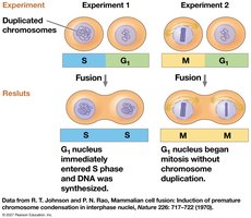
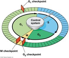
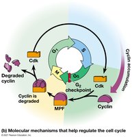
The cell cycle is regulated by a molecular control system involving cyclins and cyclin-dependent kinases (Cdks). The frequency of cell division varies with cell type and is controlled at specific checkpoints.
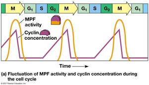
Cyclins: Proteins with cyclically fluctuating concentrations.
Cyclin-dependent kinases (Cdks): Enzymes that must be attached to a cyclin to be active.
MPF (Maturation-promoting factor): Cyclin-Cdk complex that triggers passage past the G2 checkpoint into M phase.
Cell Cycle Checkpoints
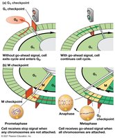
Checkpoints are control points where the cell cycle can be stopped until certain conditions are met. The G1 checkpoint is particularly important, determining whether a cell will proceed with division or enter a nondividing state (G0 phase).
G1 checkpoint: Restriction point; most important for many cells.
G0 phase: Nondividing state entered if the cell does not receive a go-ahead signal.
M checkpoint: Ensures all chromosomes are properly attached to the spindle before anaphase.
Internal and External Signals
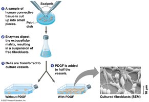
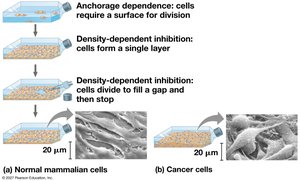
Cell division is regulated by internal surveillance mechanisms and external factors such as growth factors. Platelet-derived growth factor (PDGF) stimulates cell division in fibroblasts. Density-dependent inhibition and anchorage dependence regulate cell growth.
Growth factors: Chemical signals that stimulate cell division.
Density-dependent inhibition: Cells stop dividing when crowded.
Anchorage dependence: Cells must be attached to a surface to divide.
Loss of Cell Cycle Controls in Cancer Cells
Cancer and Cell Cycle Regulation
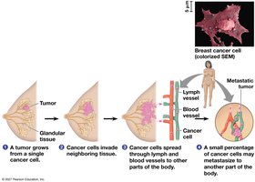
Cancer cells evade normal cell cycle controls, leading to uncontrolled division. They may produce their own growth factors, ignore external signals, or have abnormal control systems. Tumors can be benign or malignant, with malignant tumors capable of metastasis.
Transformation: Process by which cells acquire the ability to divide indefinitely.
Benign tumor: Abnormal cells remain at the original site.
Malignant tumor: Invades surrounding tissues and can spread (metastasize) to other parts of the body.
Metastasis: Spread of cancer cells to distant sites.
Scientific Skills and Summary
Interpreting Experimental Data
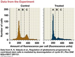
Understanding cell cycle regulation and cancer involves interpreting experimental data, such as histograms showing cell populations and fluorescence measurements.
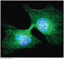
Summary of Key Concepts
The mitotic (M) phase includes prophase, prometaphase, metaphase, anaphase, telophase, and cytokinesis. Each stage is essential for accurate chromosome segregation and cell division.
Test Your Understanding
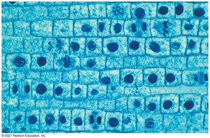
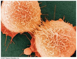
Questions and images of cells, such as those near the tip of an onion root or HeLa cancer cells, are used to assess knowledge of cell cycle concepts.
Additional info:
Equations for cell cycle regulation can be represented as:
Cell cycle checkpoints are critical for preventing errors in chromosome segregation and maintaining genomic stability.
