 Indietro
The Cell: The Fundamental Unit of Life – General Biology Study Notes
Guida di studio - Note intelligenti
 Appunti personalizzati basati sui tuoi materiali, ampliati con definizioni chiave, esempi e contesto.
The Cell: The Fundamental Unit of Life
Introduction to Microscopes
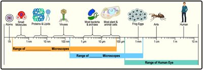
Microscopes are essential tools in biology, allowing scientists to visualize objects too small for the naked eye, such as cells and microbes. There are two main types of microscopes: light microscopes and electron microscopes, each with distinct capabilities.
Light Microscopes: Use visible light to magnify small objects, suitable for viewing most bacteria and plant/animal cells.
Electron Microscopes: Use electron beams for much higher magnification, enabling visualization of viruses, proteins, and cell structures.
Types of Electron Microscopes:
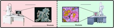
Scanning Electron Microscope (SEM): Visualizes external cell surfaces.
Transmission Electron Microscope (TEM): Visualizes internal cell structures.
Example: To measure the size of a ribosome inside a eukaryotic cell, a transmission electron microscope is required.
Prokaryotic & Eukaryotic Cells
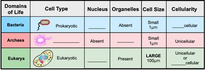
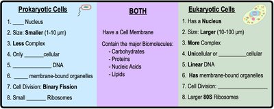
All life is classified into two broad cell types: prokaryotic and eukaryotic. These categories are fundamental to understanding cellular structure and function.
Prokaryotic Cells: Lack a nucleus and membrane-bound organelles. Includes domains Bacteria and Archaea.
Eukaryotic Cells: Possess a nucleus and various membrane-bound organelles. Includes domain Eukarya.
Example: Prokaryotic cells are found in bacteria and archaea, while eukaryotic cells are found in plants, animals, fungi, and protists.
Features of Bacterial Cells
Bacteria are the most abundant and diverse organisms on Earth. Their cellular structure is distinct from eukaryotes.
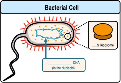
Bacterial DNA: Circular in shape, located in a region called the nucleoid.
Ribosomes: Small (70S), responsible for protein synthesis.
Cell Division: Occurs by binary fission.
Example: In bacteria, DNA is found in the nucleoid, not in a membrane-enclosed nucleus.
Features of Eukaryotic Cells
Eukaryotic cells are characterized by their complexity and presence of organelles.
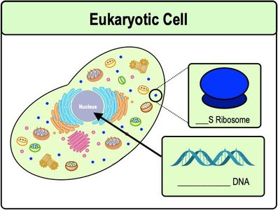
Nucleus: Packages genetic material (DNA).
DNA: Linear in shape, located inside the nucleus.
Ribosomes: Large (80S), responsible for protein synthesis.
Cell Division: Occurs by mitosis and cytokinesis.
Example: The nucleus is the organelle that packages hereditary material in eukaryotes.
Recap: Prokaryotic vs. Eukaryotic Cells
Prokaryotic and eukaryotic cells share some features but differ in key aspects.
Both: Have a cell membrane and contain major biomolecules (carbohydrates, proteins, nucleic acids, lipids).
Prokaryotes: Smaller, less complex, only unicellular, circular DNA, no membrane-bound organelles, binary fission, small ribosomes.
Eukaryotes: Larger, more complex, can be unicellular or multicellular, linear DNA, membrane-bound organelles, mitosis, large ribosomes.
Introduction to Eukaryotic Organelles
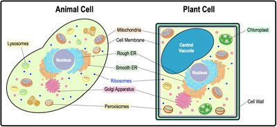
Eukaryotic cells contain several membrane-bound organelles, each with specialized functions. Animal and plant cells share many organelles, but some are unique to each type.
Animal Cells: Contain lysosomes.
Plant Cells: Contain chloroplasts and a central vacuole.
Example: Both cell types have mitochondria, nucleus, ER, Golgi apparatus, ribosomes, and peroxisomes.
Ribosomes
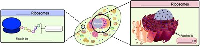
Ribosomes are non-membranous organelles responsible for protein synthesis in all living cells.
Protein Synthesis: The process conducted by ribosomes to build proteins.
Location: Ribosomes can be free-floating in the cytoplasm or attached to the rough ER.
Example: Ribosomes synthesize proteins in all cell types.
Map of Eukaryotic Organelles
Eukaryotic organelles are organized into functional groups, including the endomembrane system, energy organelles, cytoskeleton, and cell junctions.
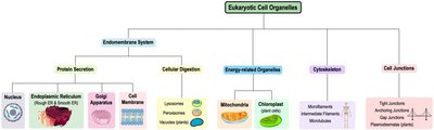
Endomembrane System: Protein Secretion
The endomembrane system is a group of membrane-bound organelles in eukaryotic cells, interconnected by vesicles. It is essential for protein secretion and cellular digestion.
Components: Nuclear envelope, endoplasmic reticulum, Golgi apparatus, transport vesicles, lysosomes, peroxisomes, vacuoles, cell membrane.
Functions: Protein secretion, cellular digestion, recycling, and transport.
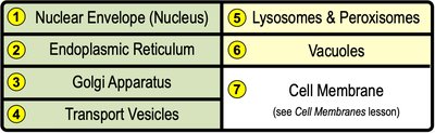
Protein Secretion Pathway
Protein secretion involves several organelles interacting in a specific sequence.
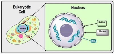
Nucleus: Stores DNA, the code for making proteins. DNA is transcribed into RNA, which is translated into protein.
Nuclear Envelope: Double membrane surrounding the nucleus, with nuclear pores for entry/exit.
Nucleolus: Assembles ribosomes and ribosomal RNA.
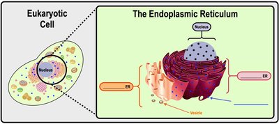
Endoplasmic Reticulum (ER)
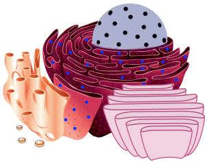
The ER is a membranous structure continuous with the nuclear envelope, with two main types:
Rough ER: Ribosome-coated surface, closer to the nucleus. Newly built proteins fold and are modified in the rER lumen.
Smooth ER: Ribosome-free surface, further from the nucleus. Synthesizes lipids and detoxifies drugs/poisons.
Golgi Apparatus
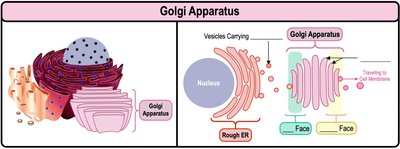
The Golgi apparatus is a stack of flat, membranous sacs functioning as a processing center for molecules synthesized in the ER.
Functions: Receives vesicles, modifies contents, repackages for export.
Ends: Receiving end is the "cis" face; shipping end is the "trans" face.
Example: Proteins are transported from rough ER → Golgi → transport vesicle → cell membrane.
Endomembrane System: Digestive Organelles
Some organelles specialize in cellular digestion and recycling.
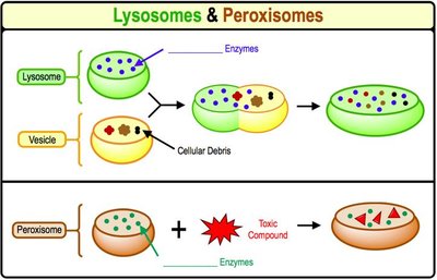
Lysosomes: Acidic vesicles containing digestive enzymes, breakdown and recycle food, debris, bacteria. Primarily found in animal cells, originate at the Golgi apparatus.
Peroxisomes: Vesicles of enzymes that breakdown toxic compounds (e.g., H2O2) and fatty acids. Found in all eukaryotic cells, originate at the rough ER.
Central Vacuole
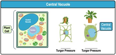
The central vacuole is a large membrane-enclosed vesicle in plant cells that degrades and recycles molecules, and helps maintain turgor pressure.
Functions: Storage, degradation, recycling, water regulation.
Turgor Pressure: Water-filled vacuole exerts pressure against the cell membrane, supporting plant structure.
Mitochondria & Chloroplasts
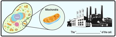
Mitochondria and chloroplasts are energy-producing organelles in eukaryotic cells.
Mitochondria
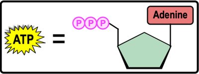
Mitochondria synthesize ATP, the cell's energy currency, through cellular respiration.
ATP (Adenosine Triphosphate): High-energy molecule used to power cellular reactions.
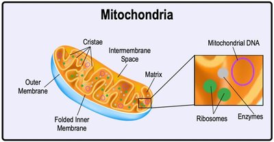
Structure: Two membranes (outer and folded inner), intermembrane space, matrix (contains enzymes, ribosomes, mitochondrial DNA).
Example: Cellular respiration is highly associated with mitochondria.
Chloroplasts
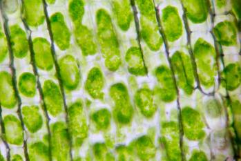
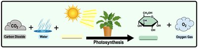
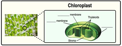
Chloroplasts are green organelles in plant cells, functioning as the site of photosynthesis.
Photosynthesis: Uses energy from sunlight to synthesize sugars (glucose).
Structure: Two membranes (outer and inner), thylakoids (disk-shaped sacs), grana (stacks of thylakoids), stroma (innermost region with enzymes, ribosomes, chloroplast DNA).
Example: The products of photosynthesis are oxygen and sugar.
Endosymbiotic Theory
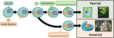
The endosymbiotic theory proposes that mitochondria and chloroplasts were once independently living prokaryotes, engulfed by a host cell to form a symbiotic relationship.
Supporting Evidence: Both organelles have small circular DNA, 70S ribosomes, replicate via binary fission, and possess double membranes.
Example: Non-nuclear DNA is found in mitochondria and chloroplasts.
Introduction to the Cytoskeleton
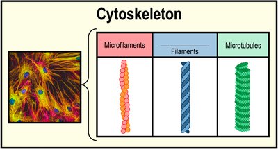
The cytoskeleton is a network of elongated proteins in the cytoplasm, providing cell shape, structure, movement, transportation, and biosignaling.
Components:
Microfilaments: Small, made of actin proteins.
Intermediate Filaments: Medium size, variable proteins.
Microtubules: Large, tube-shaped, made of tubulin proteins.
Example: Motor proteins use microtubules to transport vesicles.
Cilia & Flagella
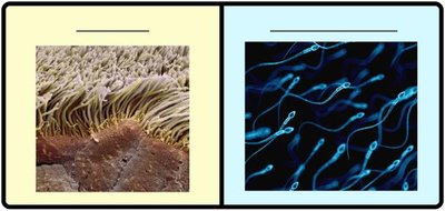
Microtubules are major structural components of cilia and flagella, which provide cell movement.
Cilia: Multiple hair-like structures that move like oars.
Flagella: Single tail-like structure that moves like a whip.
Example: In human cells, flagella move the cell, cilia move objects relative to the cell.
Cell Junctions
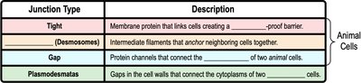
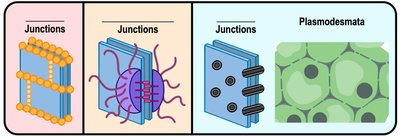
Cell junctions are structures that allow neighboring cells in eukaryotic organisms to directly communicate and link together.
Tight Junctions: Create a waterproof barrier.
Desmosomes: Anchor neighboring cells together.
Gap Junctions: Connect cytoplasm of two animal cells.
Plasmodesmata: Gaps in plant cell walls connecting cytoplasm of two plant cells.
Biological Membranes
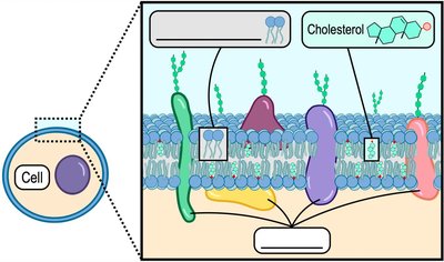
Biological membranes are primarily composed of amphipathic phospholipids, forming a bilayer with embedded proteins and cholesterol. The fluid mosaic model describes membranes as dynamic structures with proteins moving laterally within the bilayer.
Components: Phospholipids, proteins, cholesterol.
Fluid Mosaic Model: Membranes are fluid and contain a mosaic of proteins.
Types of Membrane Proteins
Membrane-associated proteins are classified as integral or peripheral, each with distinct functions.
Integral Membrane Proteins: Embedded within the membrane, often spanning the bilayer.
Peripheral Membrane Proteins: Located on the perimeter of the membrane.
Functions: Signal transduction, anchoring cytoskeleton, cell recognition, transport.
Concentration Gradients & Diffusion
A concentration gradient is a difference in the concentration of a substance between two areas. Diffusion is the movement of molecules from high to low concentration, down their gradient.
Example: Dye diffusing in water moves from high to low concentration.
Introduction to Membrane Transport
Biological membranes are selectively permeable, allowing certain molecules to cross while blocking others. Some molecules diffuse freely, others require protein facilitation.
Example: Oxygen (O2) diffuses easily across membranes; ions and large molecules require transport proteins.
Passive vs. Active Transport
There are two general types of molecular transport across membranes: passive (no energy required) and active (requires energy).
Passive Transport: Moves molecules from high to low concentration (down gradient).
Active Transport: Moves molecules from low to high concentration (up gradient), requires ATP.
Classes of Membrane Transport Proteins
Transport proteins are classified by their operation:
Uniporters: Transport one molecule at a time in one direction.
Symporters: Cotransport two or more molecules in the same direction.
Antiporters: Cotransport two or more molecules in opposite directions.
Osmosis
Osmosis is the passive diffusion of water across a semi-permeable membrane. The direction of water flow depends on the relative concentration of solutes (tonicity).
Hypotonic: Lower solute concentration.
Isotonic: Equal solute concentration.
Hypertonic: Higher solute concentration.
Water moves from hypotonic to hypertonic solutions.
Simple & Facilitated Diffusion
Simple diffusion is the direct movement of small, uncharged molecules through the membrane. Facilitated diffusion is the movement of charged or larger molecules via transport proteins, without energy expenditure.
Porins/Channels: Form membrane-spanning pores (e.g., aquaporins for water).
Transporters/Carriers: Undergo conformational changes to move molecules.
Example: Charged ions (e.g., Ca2+, Na+) require facilitated diffusion.
Active Transport
Active transport moves molecules against their concentration gradient, requiring energy (usually ATP).
Primary Active Transport: Directly driven by ATP hydrolysis (e.g., Na+/K+ pump).
Secondary Active Transport: Driven by concentration gradients established by primary active transport.
Example: Sodium-potassium pump transports 3 Na+ out and 2 K+ in, using ATP.
Endocytosis & Exocytosis
Large biomolecules are transported across cell membranes via endocytosis (entry) and exocytosis (exit).
Endocytosis: Macromolecule engulfment by the cell membrane, forming vesicles.
Types: Phagocytosis (cell "eating"), pinocytosis (cell "drinking"), receptor-mediated endocytosis (selective).
Exocytosis: Vesicle fusion with cell membrane, releasing contents to extracellular space.
Additional info: This study guide covers all major concepts from Chapter 3 of General Biology, including cell structure, organelles, membrane transport, and energy organelles, with relevant images to reinforce understanding.
