 Indietro
The Cellular Basis of Reproduction and Inheritance
Guida di studio - Note intelligenti
 Appunti personalizzati basati sui tuoi materiali, ampliati con definizioni chiave, esempi e contesto.
Chapter 8: The Cellular Basis of Reproduction and Inheritance
Introduction to Cellular Division
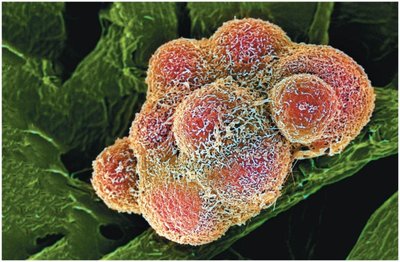
Cell division is a fundamental process in all living organisms, essential for growth, development, maintenance, and reproduction. While uncontrolled cell division leads to cancer, regulated cell division is vital for life.
Normal cell division allows organisms to grow, repair tissues, and reproduce.
Cancer cells are characterized by uncontrolled cell division, which is harmful to the organism.
Cell Division and Reproduction
Asexual and Sexual Reproduction
Organisms reproduce either asexually or sexually, each with distinct genetic outcomes.
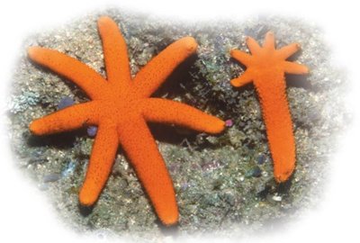
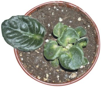
Asexual reproduction produces offspring genetically identical to the parent (clones).
Examples: Fragmentation and regeneration in sea stars, and plant cuttings that root to form new plants.
Sexual reproduction involves the fusion of gametes, resulting in genetically unique offspring due to the combination of genes from two parents.
Importance of Cell Division in Multicellular Organisms
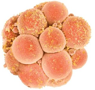
Cell division is crucial for the growth and maintenance of multicellular organisms.
Development from a single-celled zygote to a complex organism with trillions of cells.
Replacement of damaged or lost cells occurs continuously.
Cell Division in Prokaryotic Cells
Binary Fission
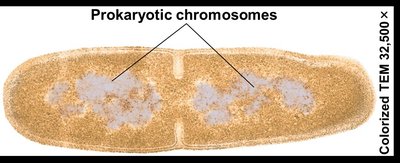
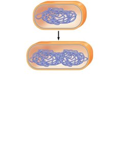
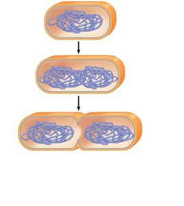
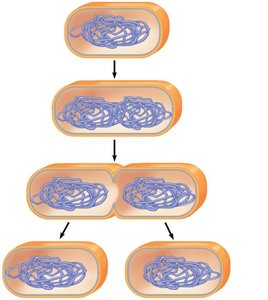
Prokaryotes, such as bacteria, reproduce by binary fission, a process that results in two genetically identical daughter cells.
Binary fission involves chromosome duplication, elongation of the cell, and division into two cells.
Prokaryotic chromosomes are typically circular and located in the cytoplasm.
The Eukaryotic Cell Cycle and Mitosis
Chromosomes and Chromatin
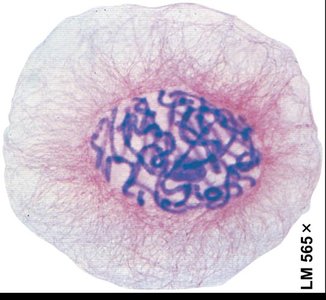
Eukaryotic cells contain multiple linear chromosomes housed within the nucleus. Each chromosome consists of a long DNA molecule and associated proteins, forming chromatin.
Chromosomes are visible only during cell division when chromatin condenses.
Chromosome Duplication and the Cell Cycle
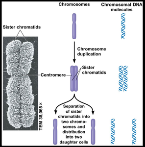
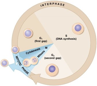
Before division, chromosomes duplicate, forming sister chromatids joined at the centromere. The cell cycle is an ordered sequence of events from one cell division to the next.
The cell cycle consists of interphase (G1, S, G2) and the mitotic phase (mitosis and cytokinesis).
Stages of Mitosis
Mitosis is divided into five main stages, each characterized by specific events involving chromosomes and the mitotic spindle.
Prophase: Chromatin condenses into visible chromosomes; spindle begins to form.
Prometaphase: Nuclear envelope breaks down; spindle fibers attach to kinetochores.
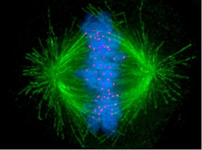
Metaphase: Chromosomes align at the metaphase plate.
Anaphase: Sister chromatids separate and move to opposite poles.
Telophase: Nuclear envelopes reform; chromosomes decondense; cytokinesis overlaps.
Interphase and Prophase
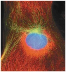
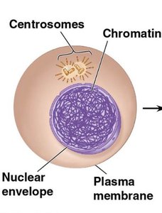
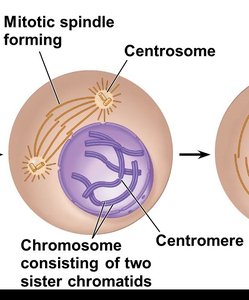
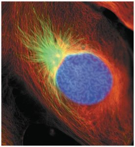
During interphase, chromosomes are duplicated and appear as loosely packed chromatin. In prophase, chromosomes condense and the mitotic spindle forms.
Control of Cell Division
Regulation of the Cell Cycle
Cell division is tightly regulated to ensure proper growth and maintenance. Several mechanisms control when and where cells divide.
Anchorage dependence: Cells must be attached to a solid surface to divide.
Density-dependent inhibition: Crowding inhibits further cell division.
Growth factors: Chemical signals that trigger cell division by binding to specific receptors.
Checkpoints: Critical points in the cell cycle regulated by growth factors.
G0 phase: Non-dividing state for cells like mature nerve and muscle cells.
Cancer and Loss of Cell Cycle Control
Cancer cells ignore normal regulatory signals, dividing uncontrollably to form tumors. Malignant tumors can invade other tissues (metastasis).
Meiosis: Reduction Division
Chromosome Number and Homologous Chromosomes
Somatic cells are diploid (2n), containing pairs of homologous chromosomes. Gametes are haploid (n), containing a single set of chromosomes. Sexual life cycles alternate between diploid and haploid stages.
Humans have 46 chromosomes (23 pairs).
Homologous chromosomes carry genes for the same traits at the same loci.
Overview of Meiosis
Meiosis consists of two consecutive divisions (Meiosis I and II) following chromosome duplication, resulting in four haploid cells.
Meiosis I: Homologous chromosomes separate, producing two haploid cells.
Meiosis II: Sister chromatids separate, resulting in four haploid cells.
Crossing over: Exchange of genetic material between homologous chromosomes during Prophase I increases genetic diversity.
Comparison of Mitosis and Meiosis
Mitosis produces two genetically identical diploid cells, while meiosis produces four genetically unique haploid cells. Key differences include pairing of homologous chromosomes, crossing over, and reduction of chromosome number in meiosis.
Feature | Mitosis | Meiosis |
|---|---|---|
Number of divisions | 1 | 2 |
Number of daughter cells | 2 | 4 |
Genetic identity | Identical | Unique |
Chromosome number | Diploid (2n) | Haploid (n) |
Role | Growth, repair | Gamete production |
Sources of Genetic Variation
Independent Assortment and Crossing Over
Genetic variation arises from independent assortment of chromosomes during meiosis and crossing over between homologous chromosomes.
The number of possible gamete combinations is calculated as , where n is the number of homologous pairs.
For humans, possible combinations per gamete.
Random fertilization further increases genetic diversity, resulting in over 70 trillion possible zygote combinations.
Crossing Over
Crossing over during Prophase I of meiosis results in recombinant chromosomes, further increasing genetic diversity among offspring.
Alterations of Chromosome Number and Structure
Nondisjunction
Nondisjunction is the failure of homologous chromosomes or sister chromatids to separate properly during meiosis, leading to abnormal chromosome numbers in gametes and offspring.
Trisomy 21 (Down syndrome): Caused by an extra chromosome 21.
Sex chromosome nondisjunction: Can result in syndromes such as Klinefelter (XXY), Turner (XO), and others.
Polyploidy: Organisms with extra sets of chromosomes, common in plants and some animals.
Sex Chromosomes | Syndrome | Frequency |
|---|---|---|
XXY | Klinefelter (male) | 1 / 2,000 |
XYY | Normal male | 1 / 2,000 |
XXX | Normal female | 1 / 1,000 |
XO | Turner (female) | 1 / 5,000 |
Alterations of Chromosome Structure
Changes in chromosome structure, such as deletions, duplications, inversions, and translocations, can cause genetic disorders and cancers. For example, chronic myelogenous leukemia (CML) is caused by a reciprocal translocation between chromosomes 9 and 22.
Additional info: These notes provide a comprehensive overview of the cellular basis of reproduction and inheritance, including the mechanisms of mitosis and meiosis, sources of genetic variation, and the consequences of chromosomal abnormalities. Understanding these processes is fundamental to genetics, development, and the study of hereditary diseases.
