 Indietro
The Cellular Basis of Reproduction and Inheritance
Guida di studio - Note intelligenti
 Appunti personalizzati basati sui tuoi materiali, ampliati con definizioni chiave, esempi e contesto.
Chapter 8: The Cellular Basis of Reproduction and Inheritance
Introduction to Cell Division and Reproduction
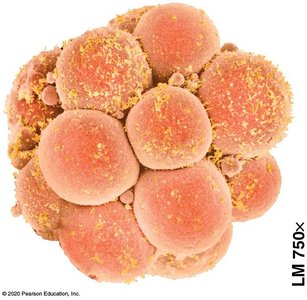
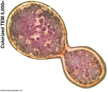
Cell division is a fundamental process by which new cells are produced from existing cells. It is essential for growth, development, tissue repair, and reproduction in all living organisms. There are two main types of reproduction: asexual, which produces genetically identical offspring from a single parent, and sexual, which combines genetic information from two parents to produce genetically varied offspring.
Asexual reproduction: Offspring are clones of the parent, inheriting identical genetic material.
Sexual reproduction: Involves the fusion of gametes (egg and sperm), resulting in genetic diversity among offspring.
Multicellular organisms: Use cell division for development, growth, and tissue repair.
Chromosome Structure and Duplication
Chromatin, Chromosomes, and Sister Chromatids
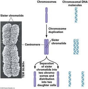
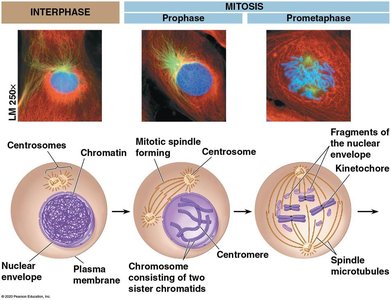
In eukaryotic cells, DNA is packaged with proteins to form chromatin, which condenses into visible chromosomes during cell division. Each duplicated chromosome consists of two identical sister chromatids joined at a region called the centromere. During cell division, sister chromatids separate, ensuring each daughter cell receives a complete set of chromosomes.
Chromatin: DNA-protein complex that condenses to form chromosomes.
Sister chromatids: Identical copies of a chromosome, joined at the centromere after DNA replication.
Chromosome counting: A duplicated chromosome has two DNA molecules; after separation, each chromatid is considered a chromosome.
The Cell Cycle
Phases of the Cell Cycle
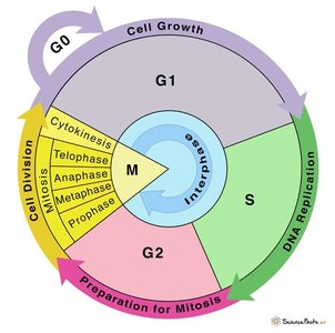
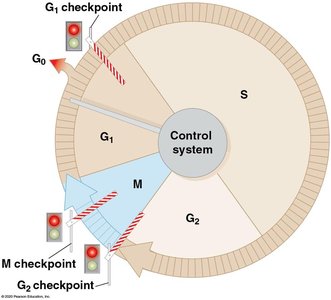
The cell cycle is the ordered sequence of events that extends from the formation of a cell to its own division into two daughter cells. It consists of interphase (G1, S, G2) and the mitotic (M) phase. Most cells spend the majority of their time in interphase, performing their normal functions and preparing for division.
G0 phase: Non-dividing state where cells perform their specialized functions.
G1 phase: Cell grows and synthesizes proteins necessary for DNA replication.
S phase: DNA is replicated, resulting in duplicated chromosomes.
G2 phase: Further growth and preparation for mitosis, including synthesis of proteins for chromosome sorting.
M phase: Includes mitosis (nuclear division) and cytokinesis (cytoplasmic division).
Regulation of the Cell Cycle
Growth Factors and Cell-Cycle Checkpoints
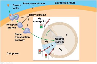
Cell division is tightly regulated by internal and external signals. Growth factors are external signals that bind to cell surface receptors and activate signal transduction pathways, leading to progression through the cell cycle. Checkpoints at G1, G2, and M phases ensure that the cell only proceeds to the next stage if conditions are favorable and the previous steps have been completed correctly.
Signal transduction: Growth factors trigger a cascade of relay proteins that activate the cell-cycle control system.
Checkpoints: Regulatory points where the cell assesses whether to continue division. The G1 checkpoint is especially important; cells that do not pass this checkpoint may enter G0.
Mitosis: Mechanism and Phases
Stages of Mitosis
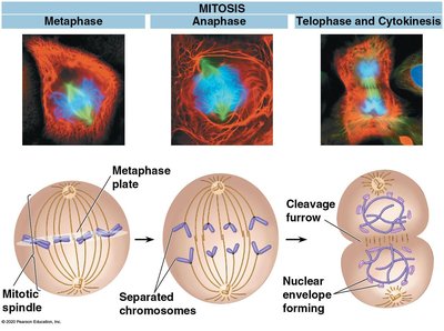
Mitosis is the process by which a eukaryotic cell divides its nucleus and distributes duplicated chromosomes into two identical daughter nuclei. It is followed by cytokinesis, which divides the cytoplasm. The stages of mitosis are:
Prophase: Chromatin condenses into chromosomes; spindle microtubules begin to form from centrosomes.
Prometaphase: Nuclear envelope breaks down; spindle fibers attach to kinetochores at centromeres.
Metaphase: Chromosomes align at the metaphase plate (cell equator).
Anaphase: Sister chromatids separate and move toward opposite poles.
Telophase: Nuclear envelopes reform around chromosomes, which decondense.
Cytokinesis: Division of the cytoplasm, resulting in two daughter cells.
Karyotypes and Chromosome Analysis
Karyotype Preparation and Interpretation
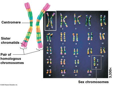
A karyotype is an ordered display of an individual's chromosomes, arranged in pairs by size, shape, and banding pattern. Karyotyping is used to detect chromosomal abnormalities and determine chromosome number and structure.
Preparation: White blood cells are stimulated to divide, arrested at metaphase, stained, photographed, and arranged into pairs.
Interpretation: Chromosome number, size, shape, and banding patterns are analyzed to identify abnormalities.
Human genome: Contains approximately 20,000 genes distributed across 23 pairs of chromosomes.
Meiosis and Sexual Life Cycles
Haploid and Diploid Stages
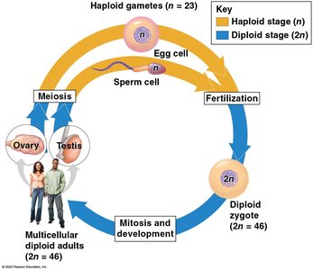
Meiosis is a specialized type of cell division that reduces the chromosome number by half, producing haploid gametes (sperm and egg). Fertilization restores the diploid chromosome number in the zygote. This alternation between haploid and diploid stages is fundamental to sexual reproduction.
Diploid (2n): Two sets of chromosomes (e.g., human somatic cells have 46 chromosomes).
Haploid (n): One set of chromosomes (e.g., human gametes have 23 chromosomes).
Meiosis: Reduces chromosome number from diploid to haploid.
Fertilization: Fusion of two haploid gametes restores diploid state.
Mechanisms of Meiosis
Meiosis I: Homologous Chromosomes Separate
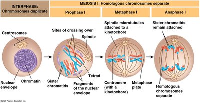
Meiosis consists of two consecutive divisions: meiosis I and meiosis II. In meiosis I, homologous chromosomes pair up and separate, reducing the chromosome number by half. Crossing over between nonsister chromatids during prophase I increases genetic diversity.
Prophase I: Homologous chromosomes pair as tetrads; crossing over occurs between nonsister chromatids.
Metaphase I: Homologous pairs align at the cell equator.
Anaphase I: Homologous chromosomes separate to opposite poles; sister chromatids remain attached.
Meiosis II: Sister Chromatids Separate
Meiosis II resembles mitosis, where sister chromatids of each chromosome are separated into different cells. No DNA replication occurs between meiosis I and II. The result is four haploid cells, each genetically distinct.
Metaphase II: Chromosomes align singly at the metaphase plate.
Anaphase II: Sister chromatids separate and move to opposite poles.
Telophase II and cytokinesis: Nuclei reform and cytoplasm divides, producing four haploid cells.
Genetic Variation in Meiosis
Crossing Over and Genetic Recombination
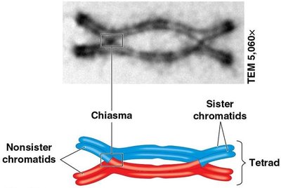
Crossing over during prophase I of meiosis results in the exchange of genetic material between nonsister chromatids, producing recombinant chromosomes with new combinations of maternal and paternal genes. This process, along with independent assortment and random fertilization, is a major source of genetic diversity in sexually reproducing organisms.
Chiasmata: Sites where crossing over occurs and chromatids remain connected.
Recombinant chromosomes: Chromosomes with new combinations of alleles.
Independent Assortment and Random Fertilization
During metaphase I, each homologous pair of chromosomes aligns independently, resulting in a variety of possible chromosome combinations in gametes. In humans, independent assortment alone allows for 223 (over 8 million) possible combinations per gamete, not including the additional variation from crossing over and random fertilization.
Independent assortment: Random orientation of homologous pairs during metaphase I.
Random fertilization: Any sperm can fertilize any egg, further increasing genetic diversity.
Errors in Meiosis and Chromosome Number
Nondisjunction and Chromosomal Disorders
Nondisjunction is the failure of homologous chromosomes or sister chromatids to separate properly during meiosis, resulting in gametes with abnormal chromosome numbers. Fertilization involving such gametes can lead to disorders such as Down syndrome (trisomy 21).
Nondisjunction: Can occur in meiosis I or II, leading to gametes with n+1 or n−1 chromosomes.
Trisomy 21 (Down syndrome): Caused by an extra copy of chromosome 21.
Comparison of Mitosis and Meiosis
Key Differences
Mitosis and meiosis are both preceded by one round of DNA replication, but differ in their outcomes and roles. Mitosis produces two genetically identical diploid cells for growth and repair, while meiosis produces four genetically distinct haploid gametes for sexual reproduction.
Feature | Mitosis | Meiosis |
|---|---|---|
Number of divisions | One | Two |
Number of daughter cells | Two | Four |
Genetic composition | Identical to parent | Genetically unique |
Chromosome number | Diploid (2n) | Haploid (n) |
Function | Growth, repair, asexual reproduction | Sexual reproduction |
Additional info: The above notes synthesize and expand upon the provided textbook content, ensuring a comprehensive and self-contained study guide for exam preparation in General Biology.
