 Indietro
The Cytoskeleton and Microscopy: Structure, Function, and Visualization in Cells
Guida di studio - Note intelligenti
 Appunti personalizzati basati sui tuoi materiali, ampliati con definizioni chiave, esempi e contesto.
The Cytoskeleton
Overview of the Cytoskeleton
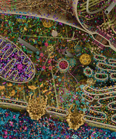
The cytoskeleton is a dynamic network of protein filaments that provides structural support, organization, and motility to cells. It is essential for maintaining cell shape, enabling intracellular transport, and facilitating cell division.
Cytoskeleton Structure: The cytoskeleton consists of three main types of filaments: microtubules, microfilaments, and intermediate filaments.
Dynamic Nature: The cytoskeleton is constantly remodeled, allowing cells to adapt to changing conditions.
Attachment: Most cellular components are anchored to the cytoskeleton.
Types of Cytoskeletal Filaments
Each filament type has distinct structural and functional properties.
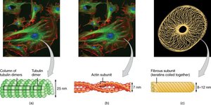
Microtubules: Hollow tubes composed of tubulin dimers. They serve as tracks for motor proteins and are crucial for cell division.
Microfilaments (Actin Filaments): Thin, flexible filaments made of actin. They are involved in cell movement, muscle contraction, and structural support.
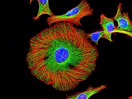
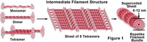
Intermediate Filaments: Rope-like filaments that provide mechanical stability and resistance to stress.
Intermediate Filaments
Intermediate filaments are the most stable cytoskeletal elements, providing structural integrity and protection against mechanical stress.
Stability: Generally remain intact but can be disassembled and repurposed.
Function: Prevent mechanical damage and maintain cell shape.
Microfilaments (Actin and Myosin)
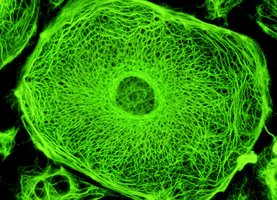
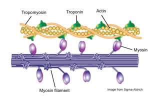
Microfilaments are involved in cell motility, muscle contraction, and structural support. Actin and myosin interact to produce movement.
Actin: Forms the core of microfilaments, enabling cell movement and shape changes.
Myosin: Motor protein that interacts with actin for muscle contraction and other motile processes.
Applications: Found in muscles, flagella, and other motile structures.
Microtubules
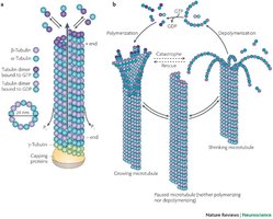
Microtubules are highly dynamic structures that are constantly assembled and disassembled. They are essential for intracellular transport and cell division.
Dynamic Instability: Microtubules undergo rapid polymerization and depolymerization.
Function: Serve as tracks for motor proteins and play a key role in mitosis.
Microscopy
Introduction to Microscopy
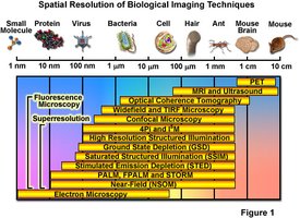
Microscopy is the technique used to visualize cells and their internal structures. It has evolved from simple light microscopes to advanced electron and fluorescence microscopes.
Resolution: Limited by the wavelength of light or electrons used.
Contrast: Enhanced by staining or specialized optics.
Living Cells: Some techniques allow observation of living cells.
Types of Light Microscopy
Light microscopy uses visible light to illuminate specimens. Different methods provide varying levels of contrast and detail.
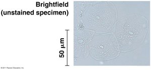
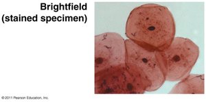
Brightfield Microscopy: Standard method; specimens can be unstained (living) or stained (dead).
Phase-Contrast Microscopy: Enhances contrast in living cells without staining.
Staining Techniques
Staining is used to increase contrast and highlight specific cellular components.
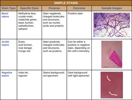
Basic Stains: Bind to negatively charged molecules (e.g., nucleic acids).
Acidic Stains: Bind to positively charged molecules (e.g., proteins).
Negative Stains: Stain the background, leaving cells unstained.
Stain Type | Specific Dyes | Purpose | Outcome | Sample Images |
|---|---|---|---|---|
Basic stains | Methylene blue, crystal violet, malachite green, basic fuchsin, carbolfuchsin, safranin | Stain negatively charged molecules and structures, such as nucleic acids and proteins | Positive stain | Cell structures highlighted |
Acidic stains | Eosin, acid fuchsin, rose bengal, Congo red | Stain positively charged molecules and structures, such as proteins | Can be either a positive or negative stain, depending on the cell's chemistry | Cell structures highlighted |
Negative stains | India ink, nigrosin | Stains background, not specimen | Dark background with light specimen | Cell outline visible |
Confocal and Fluorescence Microscopy
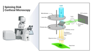
Confocal microscopy uses lasers and pinholes to produce high-resolution images of fluorescently labeled specimens. Fluorescence microscopy allows visualization of specific proteins and structures using fluorescent dyes or proteins.
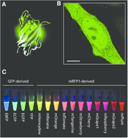
Fluorescent Proteins: Green Fluorescent Protein (GFP) is used to tag proteins for visualization.
Applications: Enables study of living cells and dynamic processes.
Green Fluorescent Protein (GFP)
Tracking Proteins with GFP
GFP is a protein originally found in jellyfish that fluoresces green under specific light. It is used as a molecular tag to visualize proteins and cellular structures in living and preserved cells.
Visualization: Requires a microscope with fluorescent filters.
Applications: Used to track protein movement and localization.
Electron Microscopy
Principles of Electron Microscopy
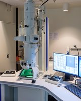
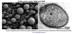
Electron microscopy uses electron beams instead of light to achieve much higher resolution. It is used to study cell surfaces and internal structures in great detail.
Scanning Electron Microscopy (SEM): Visualizes cell surfaces.
Transmission Electron Microscopy (TEM): Reveals internal cell structures.
Specimen Preparation: Requires preserved (dead) specimens.
Historical Microscopy and Cell Discovery
Robert Hooke and Early Microscopy
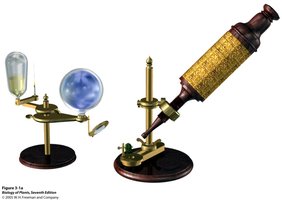
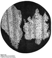
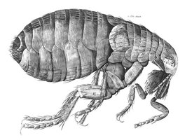
Robert Hooke was a pioneer in microscopy, using simple microscopes to observe and describe cells. He coined the term "cell" after observing cork tissue.
Micrographia: Hooke's book documenting his observations.
Cell Discovery: Cork cells appeared as "little rooms" under the microscope.

Summary Table: Cytoskeletal Filaments
Filament Type | Structure | Function | Examples |
|---|---|---|---|
Microtubules | Hollow tubes of tubulin | Cell division, transport, structure | Mitotic spindle, cilia, flagella |
Microfilaments | Thin filaments of actin | Motility, shape, contraction | Muscle fibers, cell cortex |
Intermediate Filaments | Rope-like, stable | Mechanical stability | Keratin, nuclear lamina |
Additional info: Expanded explanations and context were added to clarify cytoskeletal functions, microscopy techniques, and historical discoveries for exam preparation.
