 Indietro
The Cytoskeleton: Structure and Function Inside the Cell
Guida di studio - Note intelligenti
 Appunti personalizzati basati sui tuoi materiali, ampliati con definizioni chiave, esempi e contesto.
Inside the Cell: The Cytoskeleton
Overview of the Cytoskeleton
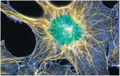
The cytoskeleton is a dynamic, complex network of protein filaments that provides structural support, facilitates cell movement, and organizes the cell's internal components. It consists of three main types of polymers: actin filaments (microfilaments), microtubules, and intermediate filaments. These elements are essential for maintaining cell shape, enabling intracellular transport, and driving cellular processes such as division and locomotion.
Actin filaments: Thinnest, involved in cell shape and movement.
Microtubules: Largest, provide tracks for organelle movement and are crucial for cell division.
Intermediate filaments: Tough, durable, provide mechanical strength.
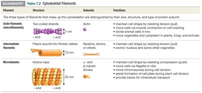
Summary Table: Cytoskeletal Filaments
The following table summarizes the structure, subunits, and functions of the three cytoskeletal filaments:
Filament | Structure | Subunits | Functions |
|---|---|---|---|
Actin filaments (microfilaments) | Two coiled strands | Actin | Maintain cell shape, movement, muscle contraction, cytokinesis, cytoplasmic streaming |
Intermediate filaments | Fibers wound into thicker cables | Keratin, lamins, others | Maintain cell shape, anchor organelles, nuclear envelope support |
Microtubules | Hollow tube | α- and β-tubulin dimers | Maintain cell shape, move organelles, cell division, tracks for transport |
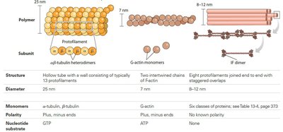
Structural Properties of Cytoskeletal Filaments
Each cytoskeletal filament has unique structural properties, subunit composition, and polarity, which determine its function in the cell.
Polymer | Subunit | Structure | Diameter | Monomers | Polarity | Nucleotide Substrate |
|---|---|---|---|---|---|---|
Microtubule | αβ-tubulin heterodimers | Hollow tube | 25 nm | α-tubulin, β-tubulin | Plus, minus ends | GTP |
Actin filament | G-actin monomers | Two intertwined chains | 7 nm | G-actin | Plus, minus ends | ATP |
Intermediate filament | IF dimer | Eight protofilaments joined end to end | 8–12 nm | Six classes of proteins | No known polarity | None |
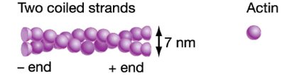
Actin Filaments
Structure of Actin Filaments
Actin filaments are the thinnest cytoskeletal polymers, composed of two twisted strands of actin subunits. They exhibit structural polarity, with a plus end (faster growth) and a minus end. Actin filaments can form bundles or cross-linked networks, contributing to cell shape and movement.
Diameter: 7 nm
Polarity: Plus and minus ends
Subunit: Actin monomer
Functions of Actin Filaments
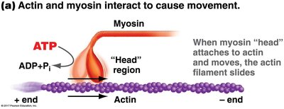
Actin filaments play a crucial role in maintaining cell shape, enabling cell movement, and facilitating muscle contraction. The motor protein myosin interacts with actin filaments, using ATP to generate force and movement.
Cell shape maintenance
Cell movement
Muscle contraction
Cytokinesis (cell division)
Cytoplasmic streaming
Actin-Myosin Interactions
Many cellular movements are based on actin–myosin interactions. Myosin uses ATP to change shape and move along actin filaments, causing processes such as cytoplasmic streaming in plants and cytokinesis in animals.
ATP hydrolysis: Provides energy for myosin movement
Sliding filament mechanism: Myosin head attaches to actin and moves, sliding the filament
Intermediate Filaments
Structure of Intermediate Filaments
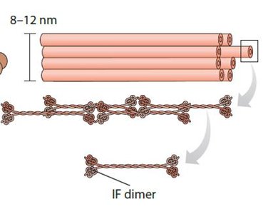
Intermediate filaments are composed of various proteins (e.g., keratin, nuclear lamins) and are formed from coiled coil dimers that overlap. They lack structural polarity and are tough and durable, providing mechanical strength to cells.
Diameter: 8–12 nm
Subunit: IF dimer
No structural polarity
Functions of Intermediate Filaments
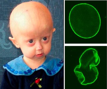
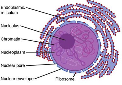
Intermediate filaments provide structural support, anchor organelles, and form the nuclear lamina, which supports the nuclear envelope. Mutations in nuclear lamins can cause diseases such as progeria, characterized by premature aging.
Keratin: Found in hair, claws, fingernails
Nuclear lamins: Support nuclear envelope
Mechanical strength: Protects cells from mechanical stress
Microtubules
Structure of Microtubules
Microtubules are the largest cytoskeletal elements, consisting of hollow tubes made of α- and β-tubulin heterodimers. They exhibit structural polarity, with plus and minus ends, and are dynamic, growing faster at their plus ends.
Diameter: 25 nm
Subunit: α- and β-tubulin heterodimers
Polarity: Plus and minus ends
Microtubule Organizing Centers (MTOCs)
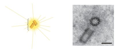
Microtubules originate from the microtubule organizing center (MTOC), which anchors the minus end. In animal cells, the MTOC is called the centrosome, containing centrioles. Plus ends grow outward, radiating throughout the cell.
Centrosome: Main MTOC in animal cells
Centrioles: Cylindrical structures within the centrosome
Functions of Microtubules
Microtubules provide structural support, facilitate organelle movement, and are essential for cell division. They serve as tracks for motor proteins such as kinesin and dynein, which transport vesicles and organelles.
Organelle stability
Cell division: Separation of chromosomes
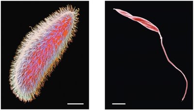
Cell locomotion: Cilia and flagella
Intracellular transport: Tracks for movement
Dynamic Instability of Microtubules
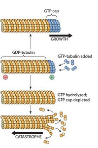
Microtubules undergo rapid shrinking and regrowth, a process known as dynamic instability. This is regulated by the addition and loss of GTP-tubulin at the plus end, leading to phases of growth and catastrophe.
GTP cap: Stabilizes growing microtubule
Catastrophe: Loss of GTP cap causes rapid depolymerization
Microtubules in Cell Division and Transport
Microtubules form the mitotic spindle during cell division, ensuring proper chromosome separation. Motor proteins such as kinesin and dynein move vesicles and organelles along microtubule tracks, using ATP for energy.
Kinesin: Moves toward plus end
Dynein: Moves toward minus end
ATP hydrolysis: Powers motor protein movement
Cilia and Flagella: Microtubule-Based Structures
Eukaryotic cilia and flagella are composed of microtubules arranged in an axoneme with a "9 + 2" structure. Dynein motor proteins cause bending by walking along adjacent microtubule doublets, enabling cell movement.
Axoneme: "9 + 2" arrangement of microtubules
Dynein: Motor protein causing bending
Prokaryotic flagella: Made of flagellin, not microtubules
Summary: Cytoskeletal Elements
The cytoskeleton is essential for cell structure, movement, and internal organization. Each filament type has distinct properties and functions, contributing to the dynamic nature of the cell.
Actin filaments: Cell shape, movement, muscle contraction
Intermediate filaments: Mechanical strength, nuclear envelope support
Microtubules: Organelle movement, cell division, cilia and flagella
Additional info: The cytoskeleton is also involved in signaling pathways and cellular responses to environmental changes, highlighting its importance in cell biology.
