 Indietro
The Cytoskeleton: Structure, Function, and Dynamics in Eukaryotic Cells
Guida di studio - Note intelligenti
 Appunti personalizzati basati sui tuoi materiali, ampliati con definizioni chiave, esempi e contesto.
The Cytoskeleton
Overview and Functions
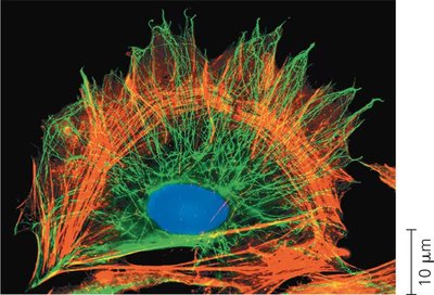
The cytoskeleton is a dynamic network of fibers that extends throughout the cytoplasm of eukaryotic cells, providing structural support, organization, and facilitating cellular activities. It is composed of three main types of fibers: microtubules, microfilaments (actin filaments), and intermediate filaments. The cytoskeleton is essential for maintaining cell shape, anchoring organelles, and enabling cell motility and intracellular transport.
Mechanical Support: Maintains cell shape, especially in animal cells lacking cell walls.
Anchorage: Provides anchorage for organelles and cytosolic enzymes.
Dynamic Remodeling: Can be rapidly assembled and disassembled, allowing changes in cell shape and movement.
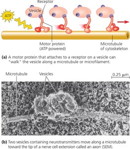
Cell Motility: Facilitates movement of cells and cell parts, often through interaction with motor proteins.
Example: Vesicles containing neurotransmitters migrate along cytoskeletal tracks to the tips of axons in nerve cells.
Components of the Cytoskeleton
Microtubules
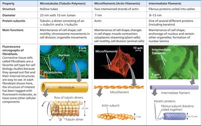
Microtubules are hollow tubes made of tubulin dimers (α-tubulin and β-tubulin). They are the thickest cytoskeletal fibers and play key roles in cell shape, organelle movement, and chromosome separation during cell division.
Structure: Hollow tubes, 25 nm diameter, composed of tubulin dimers.
Polarity: Plus and minus ends allow dynamic growth and shrinkage.
Main Functions: Maintenance of cell shape, tracks for motor proteins, chromosome movement during mitosis/meiosis.
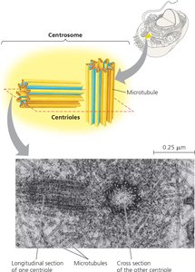
Centrosomes and Centrioles: In animal cells, microtubules originate from the centrosome, which contains a pair of centrioles arranged in a ring of nine triplets.
Cilia and Flagella
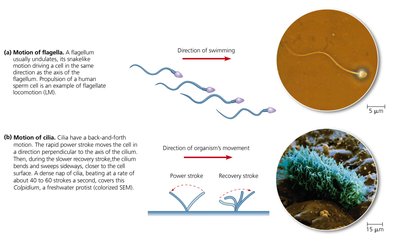
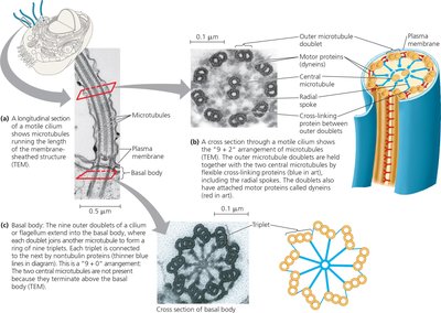
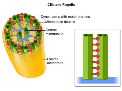
Cilia and flagella are cellular extensions containing microtubules arranged in a "9 + 2" pattern. They are responsible for cell movement and moving fluids over cell surfaces. Cilia are usually numerous and short, while flagella are longer and fewer in number.
Beating Patterns: Flagella move with an undulating motion; cilia move with alternating power and recovery strokes.
Basal Body: Anchors the microtubule assembly; structurally similar to a centriole.
Primary Cilium: Nonmotile, acts as a signal-receiving antenna for the cell.
Microfilaments (Actin Filaments)
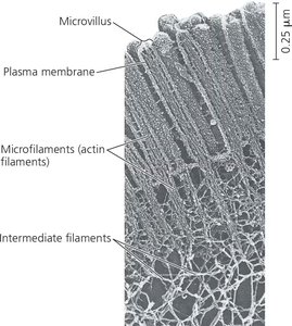
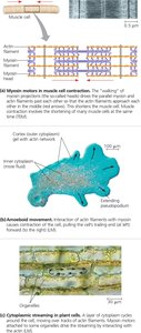
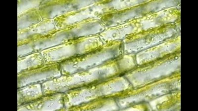
Microfilaments are thin, solid rods composed of actin. They are present in all eukaryotic cells and are specialized for bearing tension. Microfilaments support cell shape, form the cortex, and are involved in cell motility, muscle contraction, and cytoplasmic streaming.
Structure: Twisted double chain of actin subunits, 7 nm diameter.
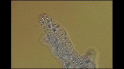
Main Functions: Maintenance of cell shape, changes in cell shape, muscle contraction, cell motility (amoeboid movement), cytoplasmic streaming.
Cortex: Network of microfilaments just inside the plasma membrane gives the cell a gel-like consistency.
Microvilli: Bundles of microfilaments reinforce cellular extensions that increase surface area.
Intermediate Filaments
Intermediate filaments are fibers with diameters between microtubules and microfilaments. They are composed of various proteins, including keratins, and are specialized for bearing tension. Intermediate filaments are more permanent structures, providing stability and anchoring organelles.
Structure: Fibrous proteins coiled into cables, 8–12 nm diameter.
Main Functions: Maintenance of cell shape, anchorage of nucleus and other organelles, formation of nuclear lamina.
Stability: More permanent than microtubules and microfilaments; persist even after cell death.
Summary Table: Cytoskeletal Elements
Property | Microtubules | Microfilaments | Intermediate Filaments |
|---|---|---|---|
Structure | Hollow tubes | Two intertwined strands of actin | Fibrous proteins coiled into cables |
Diameter | 25 nm with 15 nm lumen | 7 nm | 8–12 nm |
Protein Subunits | Tubulin (α and β) | Actin | One of several proteins (e.g., keratin) |
Main Functions | Cell shape, organelle movement, chromosome movement | Cell shape, muscle contraction, cell motility, cytoplasmic streaming | Cell shape, anchorage, nuclear lamina |
Mechanisms of Cytoskeletal Function
Motor Proteins and Cellular Movement
Cell motility and intracellular transport are achieved through the interaction of cytoskeletal elements with motor proteins. These proteins use ATP to "walk" along cytoskeletal tracks, moving vesicles and organelles to their destinations.
Example: Dynein moves along microtubules in cilia and flagella, causing bending.
Actin-Myosin Interaction: Responsible for muscle contraction and amoeboid movement.
Equation: ATP hydrolysis provides energy for motor protein movement:
Applications and Clinical Relevance
Genetic Disorders
Defects in cytoskeletal components can lead to diseases. For example, Kartagener’s syndrome results from immotile cilia and flagella due to defective dynein arms, causing infertility and respiratory issues.
Example: Immotile sperm and recurrent lung infections in affected males.
Conclusion
The cytoskeleton is a fundamental component of eukaryotic cells, providing structural integrity, facilitating movement, and organizing cellular activities. Understanding its structure and function is essential for comprehending cell biology and its implications in health and disease.
