 Indietro
The Human Circulatory System: Structure, Function, and Fetal Adaptations
Guida di studio - Note intelligenti
 Appunti personalizzati basati sui tuoi materiali, ampliati con definizioni chiave, esempi e contesto.
The Circulatory System
Overview and Function
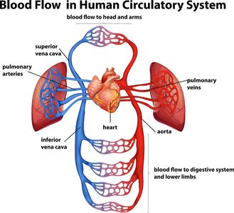
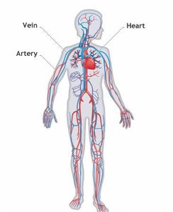
The circulatory system is an organ system responsible for the transport of fluids, nutrients, gases, immune cells, and hormones throughout the body. It consists of a network of vessels (arteries, veins, capillaries) connected to a central pump, the heart.
Key Point 1: The circulatory system enables diffusion and transport of essential substances to and from cells.
Key Point 2: Blood is the main circulating fluid, containing cells and plasma.
Example: Oxygen is transported from the lungs to tissues, while carbon dioxide is carried from tissues to the lungs for exhalation.
Blood: Components and Functions
Blood as a Connective Tissue
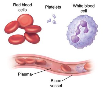
Blood is a specialized connective tissue with a fluid matrix called plasma. It contains three main cellular components: red blood cells, white blood cells, and platelets.
Key Point 1: Plasma is the fluid portion, a protein-salt solution that suspends cells, gases, and nutrients.
Key Point 2: Red blood cells (RBCs) transport oxygen (O2) and carbon dioxide (CO2).
Key Point 3: White blood cells (WBCs) are immune cells that defend against infection.
Key Point 4: Platelets are cell fragments involved in blood clotting.
Example: Platelets form clots to prevent blood loss at injury sites.
Coagulation: Blood Clot Formation
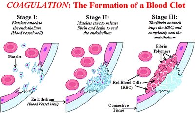
Blood clotting is a multi-step process involving platelets and fibrin to seal damaged vessels.
Key Point 1: Platelets adhere to vessel walls at injury sites.
Key Point 2: Fibrin is released to form a mesh, trapping blood cells.
Key Point 3: The clot prevents further bleeding and initiates healing.
Example: Coagulation is essential for wound healing and preventing excessive blood loss.
Interstitial Fluid
Role and Composition
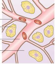
Interstitial fluid bathes and surrounds tissue cells, facilitating exchange of nutrients and waste. It is similar to plasma but contains less protein.
Key Point 1: Interstitial fluid is the medium for diffusion between blood and cells.
Key Point 2: It is essential for cellular homeostasis.
Blood Vessels: Structure and Function
Types of Blood Vessels
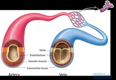
The body contains three major types of blood vessels: arteries, veins, and capillaries.
Key Point 1: Arteries carry blood away from the heart, usually oxygenated (except pulmonary artery).
Key Point 2: Veins carry blood toward the heart, usually deoxygenated (except pulmonary veins).
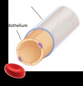
Key Point 3: Capillaries are thin vessels where gas and nutrient exchange occurs.
Vessel Wall Structure
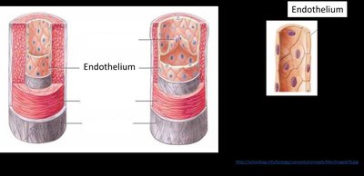
All vessels have a smooth endothelium. Arteries and veins also have smooth muscle and connective tissue layers.
Key Point 1: Arteries have thick muscle walls to withstand high pressure.
Key Point 2: Veins have thinner walls and valves to prevent backflow.
Key Point 3: Capillaries are only slightly wider than a single blood cell, facilitating exchange.
The Heart: Structure and Circulation
Four-Chambered Heart
Mammals and birds have a four-chambered heart, separating oxygenated and deoxygenated blood.
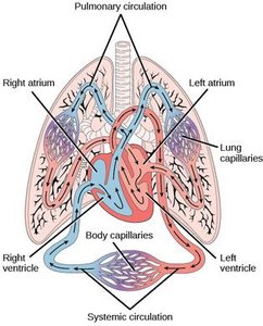
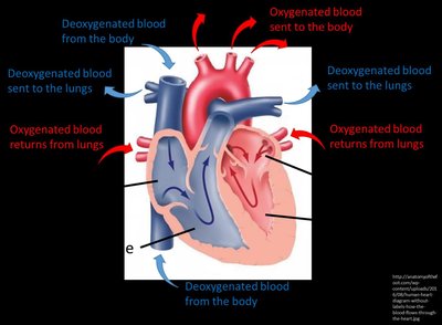
Key Point 1: Right atrium receives deoxygenated blood; right ventricle sends it to lungs.
Key Point 2: Left atrium receives oxygenated blood; left ventricle sends it to the body.
Heart Valves
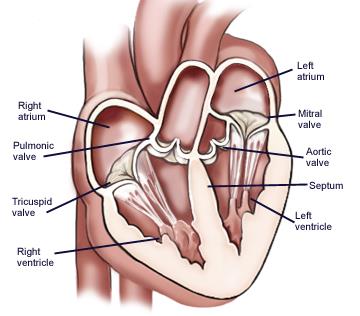
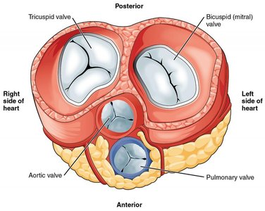
Valves prevent backflow during heart contractions, ensuring unidirectional blood flow.
Key Point 1: Major valves include tricuspid, mitral, aortic, and pulmonary valves.
Key Point 2: Valves open and close in response to pressure changes during the cardiac cycle.
Cardiac Conduction and Heart Rate
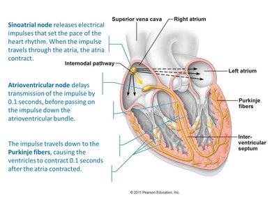
The sinoatrial (SA) node generates electrical impulses that coordinate heart contractions. The autonomic nervous system modulates heart rate.
Key Point 1: Sympathetic stimulation increases heart rate; parasympathetic decreases it.
Key Point 2: Systole is contraction; diastole is relaxation.
Formula:
Circulatory Pathways
Systemic and Pulmonary Circulation
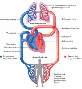
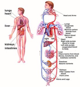
Blood flows through two main circuits: systemic (body) and pulmonary (lungs).
Key Point 1: Deoxygenated blood returns to the right heart, sent to lungs for oxygenation.
Key Point 2: Oxygenated blood returns to the left heart, sent to the body.
Key Point 3: Capillaries facilitate exchange; veins return blood to the heart.
Fetal Circulation
Adaptations for Oxygen and Nutrient Exchange
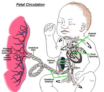
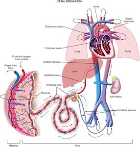
The fetal circulatory system is modified to allow oxygen and nutrients from the placenta, bypassing the lungs.
Key Point 1: The placenta exchanges gases and nutrients between mother and fetus.
Key Point 2: The umbilical vein carries oxygenated blood to the fetus; umbilical arteries return deoxygenated blood to the placenta.
Key Point 3: The oval opening (foramen ovale) and arterial duct (ductus arteriosus) allow blood to bypass the fetal lungs.
Summary Table: Blood Vessel Comparison
Structural and Functional Differences
Vessel Type | Direction of Blood Flow | Wall Structure | Valves | Oxygen Content |
|---|---|---|---|---|
Artery | Away from heart | Thick muscle, connective tissue, endothelium | No | Usually oxygenated (except pulmonary artery) |
Vein | Toward heart | Thin muscle, connective tissue, endothelium | Yes | Usually deoxygenated (except pulmonary vein) |
Capillary | Connects arteries and veins | Single layer endothelium | No | Site of gas/nutrient exchange |
Additional info:
Blood pressure is measured as systolic/diastolic values, indicating pressure during contraction and relaxation.
Fetal circulation adaptations are temporary and close after birth.
