 Indietro
lecture 22-23: The Molecular Basis of Inheritance: DNA Structure and Replication
Guida di studio - Note intelligenti
 Appunti personalizzati basati sui tuoi materiali, ampliati con definizioni chiave, esempi e contesto.
The Molecular Basis of Inheritance
DNA as the Genetic Material
DNA (deoxyribonucleic acid) is the molecule responsible for genetic inheritance in all living organisms. It is a polymer composed of nucleotides, each containing a nitrogenous base, a pentose sugar (deoxyribose), and a phosphate group. The discovery that DNA is the genetic material was supported by Erwin Chargaff's findings in 1950, which showed that the base composition of DNA varies between species, but within any species, the number of adenine (A) and thymine (T) bases are equal, as are the number of guanine (G) and cytosine (C) bases.
Nucleotide Structure: Each nucleotide consists of a phosphate group, deoxyribose sugar, and one of four nitrogenous bases: adenine, thymine, cytosine, or guanine.
Chargaff's Rules: In any species, A = T and G = C.
Species Variation: The ratio of bases varies between species, providing evidence for DNA's role in heredity.

Determining the Structural Model of DNA
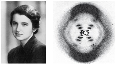
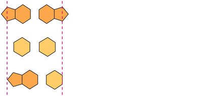
The structure of DNA was elucidated through X-ray crystallography, notably by Rosalind Franklin and Maurice Wilkins. Franklin's X-ray diffraction photograph provided critical evidence for the helical structure of DNA. Watson and Crick built on this data, proposing the double helix model in 1953. They determined that base pairing occurs between a purine and a pyrimidine, resulting in a uniform width consistent with X-ray data.
X-ray Crystallography: Revealed the helical structure of DNA.
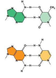
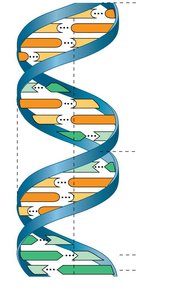
Base Pairing: Adenine pairs with thymine, and guanine pairs with cytosine.
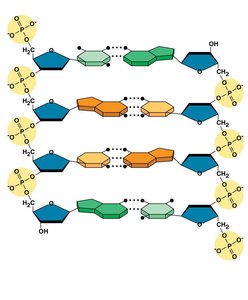
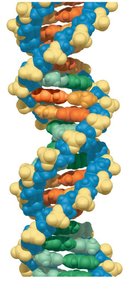
Double Helix: Two strands of DNA coil around each other, held together by hydrogen bonds between complementary bases.
Key Features of DNA Structure
The double helix model explains the physical and chemical properties of DNA. The strands are antiparallel, meaning they run in opposite directions. The backbone is formed by alternating sugar and phosphate groups, while the nitrogenous bases project inward, forming base pairs.
Antiparallel Strands: One strand runs 5' to 3', the other 3' to 5'.
Hydrogen Bonding: A-T pairs have two hydrogen bonds; G-C pairs have three.
Uniform Width: Purine-pyrimidine pairing ensures consistent diameter.
DNA Replication: The Basic Principle
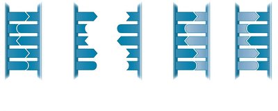
DNA replication is the process by which a cell copies its DNA before cell division. The two strands of the double helix are complementary, allowing each strand to serve as a template for the synthesis of a new strand. Replication follows the base-pairing rules, ensuring accurate transmission of genetic information.
Template Strand: Each original strand guides the formation of a new complementary strand.
Replication Mechanism: The parent molecule unwinds, and daughter strands are synthesized.


Origins of Replication
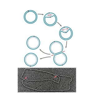
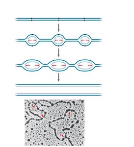
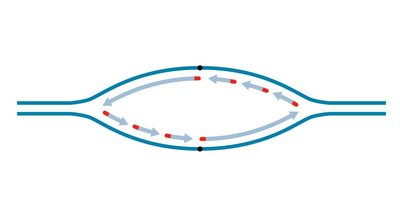
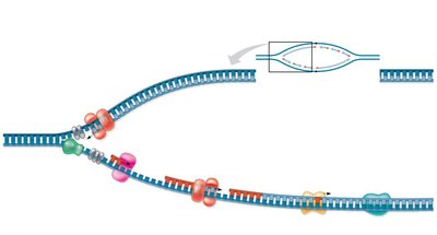
Replication begins at specific locations called origins of replication. In prokaryotes, there is typically a single origin, while eukaryotic chromosomes have multiple origins. Replication proceeds bidirectionally from each origin, forming replication bubbles.
Replication Bubble: Region where DNA strands are separated and replication occurs.
Replication Fork: Y-shaped region at the end of each bubble where new DNA is synthesized.
Enzymes and Proteins in DNA Replication
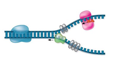
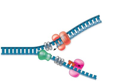
DNA replication is facilitated by a suite of enzymes and proteins, each addressing specific challenges in the process. Helicase unwinds the DNA, single-strand binding proteins stabilize the unwound strands, and topoisomerase relieves overwinding. Primase synthesizes a short RNA primer, which is required for DNA polymerase to begin synthesis.
Helicase: Unwinds the DNA double helix.
Single-Strand Binding Proteins: Stabilize separated strands.
Topoisomerase: Relieves strain ahead of the replication fork.
Primase: Synthesizes RNA primers.
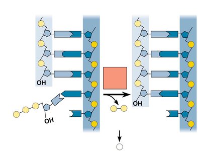
DNA Polymerase: Adds nucleotides to the 3' end of the primer.
Antiparallel Elongation and Strand Synthesis
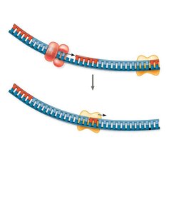
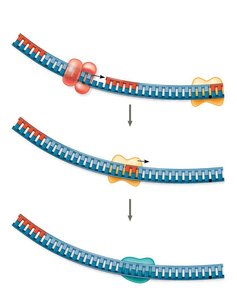
The antiparallel nature of DNA affects replication. DNA polymerases can only add nucleotides to the 3' end, so synthesis occurs in the 5' to 3' direction. The leading strand is synthesized continuously toward the replication fork, while the lagging strand is synthesized discontinuously away from the fork in short segments called Okazaki fragments.
Leading Strand: Synthesized continuously.
Lagging Strand: Synthesized in Okazaki fragments, later joined by DNA ligase.
Okazaki Fragments: Short DNA segments on the lagging strand.
DNA Ligase: Joins Okazaki fragments.
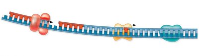
The DNA Replication Complex
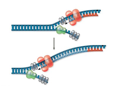
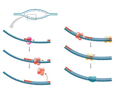
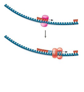
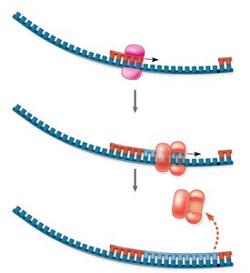
The proteins involved in DNA replication form a large complex, sometimes referred to as the "DNA replication machine." This complex may be stationary, with DNA polymerase molecules "reeling in" parental DNA and extruding newly synthesized daughter DNA.
Replication Machine: Multi-protein complex responsible for DNA synthesis.
Stationary Model: DNA moves through the complex rather than the complex moving along DNA.
Proofreading and Repairing DNA
DNA polymerases proofread newly synthesized DNA, correcting errors. Additional repair mechanisms, such as mismatch repair and nucleotide excision repair, fix errors and damage caused by environmental factors like cigarette smoke and X-rays.
Proofreading: DNA polymerase removes incorrect nucleotides.
Mismatch Repair: Repair enzymes correct base-pairing errors.
Nucleotide Excision Repair: Nuclease removes damaged DNA, which is replaced by DNA polymerase and sealed by DNA ligase.
Replicating the Ends of DNA Molecules
In eukaryotes, the linear nature of chromosomes presents a challenge for replication at the ends. DNA polymerase cannot complete the 5' ends, leading to progressive shortening. Telomeres, repetitive sequences at chromosome ends, postpone gene erosion. Telomerase extends telomeres in germ cells, with implications for aging and cancer.
Telomeres: Repetitive DNA sequences at chromosome ends.
Telomerase: Enzyme that extends telomeres in germ cells.
Biological Significance: Telomere shortening is linked to aging; telomerase activity is associated with cancer.
Chromatin Structure and DNA Packing
Eukaryotic chromosomes consist of DNA tightly packed with proteins, forming chromatin. Chromatin undergoes structural changes during the cell cycle, allowing for efficient packing and accessibility. The basic unit of chromatin is the nucleosome, a segment of DNA wrapped around histone proteins. Chromatin can be loosely packed (euchromatin) or highly condensed (heterochromatin).
Nucleosome: DNA wrapped around histone proteins.
Chromatin Fibers: 10-nm fiber (beads on a string), 30-nm fiber, looped domains, metaphase chromosome.
Euchromatin: Loosely packed, accessible for transcription.
Heterochromatin: Highly condensed, less accessible.
Summary of Key Concepts
DNA's double helix structure enables faithful replication and inheritance. Replication involves a complex interplay of enzymes and proteins, ensuring accuracy and repair. Chromatin structure allows DNA to be efficiently packed and regulated within the nucleus.
Double Helix: Structure enables complementary base pairing and replication.
Replication: Leading strand synthesized continuously; lagging strand synthesized in fragments.
Chromatin: DNA-protein complex allows packing and regulation.
