 Indietro
The Molecular Basis of Inheritance: DNA Structure and Replication
Guida di studio - Note intelligenti
 Appunti personalizzati basati sui tuoi materiali, ampliati con definizioni chiave, esempi e contesto.
Chapter 16: The Molecular Basis of Inheritance
The Big Question: How Does DNA Replication Transmit Genetic Information?
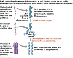
DNA replication is the process by which genetic information is faithfully transmitted from parent cells to daughter cells during cell division. This mechanism ensures that genetic traits are inherited and that the continuity of life is maintained across generations.
Griffith's Experiment and Transformation
Discovery of the Transforming Principle
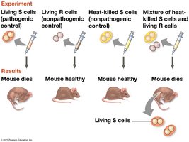
Frederick Griffith's experiments in 1928 demonstrated that a genetic trait could be transferred between bacterial strains. He worked with two strains of Streptococcus pneumoniae: a pathogenic (S) strain and a nonpathogenic (R) strain. When heat-killed S cells were mixed with living R cells and injected into mice, the mice died, and living S cells were recovered, indicating that some "transforming principle" from the dead S cells converted R cells into S cells.
Key Point: Genetic information can be transferred between organisms, suggesting a molecular basis for inheritance.
Example: Transformation of nonpathogenic bacteria into pathogenic forms.
Hershey–Chase Experiment: DNA as Genetic Material
Evidence from Bacteriophages
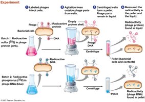
In 1952, Alfred Hershey and Martha Chase used bacteriophages (viruses that infect bacteria) to determine whether DNA or protein was the genetic material. By labeling phage DNA with radioactive phosphorus and protein with radioactive sulfur, they showed that only DNA entered bacterial cells and directed viral replication, confirming DNA as the genetic material.
Key Point: DNA, not protein, carries genetic information in viruses and cells.
Example: Radioactive DNA, but not protein, was found inside infected bacteria.
Chargaff's Rules and DNA Composition
Base Pairing and Species Variation
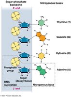
Erwin Chargaff discovered that DNA is composed of four types of nucleotides, each containing a nitrogenous base (adenine, thymine, guanine, cytosine), a sugar, and a phosphate group. He found that the amount of adenine equals thymine (A = T) and guanine equals cytosine (G = C) in any species, though the overall base composition varies between species.
Key Point: The equivalence of A to T and G to C is explained by specific base pairing in the DNA double helix.
Discovery of DNA Structure
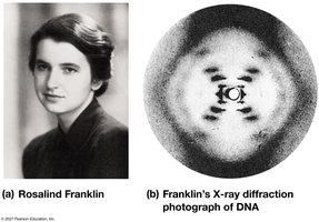
Rosalind Franklin's X-ray Diffraction
Rosalind Franklin's X-ray diffraction images provided crucial evidence that DNA is a helical molecule composed of two strands. Her work enabled Watson and Crick to deduce the double helix structure of DNA.
Key Point: X-ray diffraction patterns indicated a repeating helical structure.
The Double Helix Model
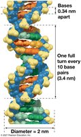
James Watson and Francis Crick proposed that DNA consists of two antiparallel strands forming a double helix, with a uniform diameter of 2 nm and one full turn every 3.4 nm. The strands are held together by specific base pairing: A pairs with T, and G pairs with C.
Key Point: The double helix explains how genetic information is stored and replicated.
Base Pairing and Hydrogen Bonds
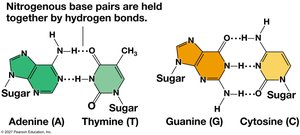
Base pairing occurs between a purine (A or G) and a pyrimidine (T or C), ensuring a uniform width. Adenine forms two hydrogen bonds with thymine, and guanine forms three hydrogen bonds with cytosine.
Key Point: Hydrogen bonds stabilize the DNA double helix and ensure accurate base pairing.
DNA Replication: Mechanisms and Models
Semiconservative Replication
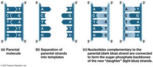
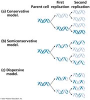
DNA replication is semiconservative: each new DNA molecule consists of one parental (old) strand and one newly synthesized strand. Competing models included conservative (parental strands rejoin) and dispersive (strands are mixtures of old and new segments).
Key Point: Complementary base pairing allows each strand to serve as a template for a new strand.
Models of Replication
The three models of DNA replication are:
Conservative: Parental strands rejoin after replication.
Semiconservative: Each daughter molecule has one old and one new strand.
Dispersive: Each strand is a mix of old and new DNA.
Meselson–Stahl Experiment
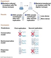
Matthew Meselson and Franklin Stahl used isotopic labeling to distinguish old and new DNA strands, providing experimental evidence for the semiconservative model of replication.
Key Point: After one replication, DNA molecules had intermediate density; after two, both intermediate and light DNA were present, supporting the semiconservative model.
DNA Replication in Cells
Origins of Replication
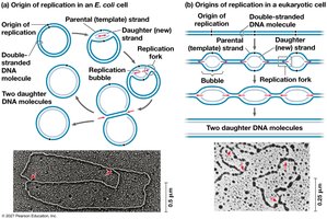
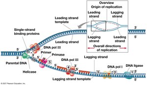
Replication begins at specific sites called origins of replication. Bacterial chromosomes typically have a single origin, while eukaryotic chromosomes have multiple origins. Replication proceeds bidirectionally from each origin, forming replication bubbles.
Key Point: Multiple origins in eukaryotes speed up the replication of large genomes.
Unwinding and Priming the DNA
At the replication fork, several proteins coordinate to unwind and stabilize the DNA:
Helicase: Unwinds the double helix.
Single-strand binding proteins: Stabilize unwound DNA.
Topoisomerase: Relieves strain ahead of the fork.
Primase: Synthesizes short RNA primers to initiate DNA synthesis.
DNA Polymerase and Synthesis
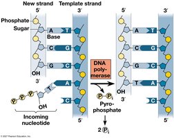
DNA polymerases add nucleotides to the 3′ end of a primer, synthesizing new DNA in the 5′ to 3′ direction. Each incoming nucleotide is a nucleoside triphosphate, which loses two phosphates as pyrophosphate during incorporation.
Key Point: DNA polymerases require a primer and a template strand to begin synthesis.
Leading and Lagging Strands
Because DNA polymerase can only synthesize in the 5′ to 3′ direction, replication is continuous on the leading strand and discontinuous on the lagging strand. The lagging strand is synthesized as short Okazaki fragments, which are later joined by DNA ligase.
Key Point: The antiparallel structure of DNA necessitates different replication strategies for each strand.
Replication Proteins
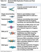
The main proteins involved in bacterial DNA replication and their functions are summarized below:
Protein | Function |
|---|---|
Helicase | Unwinds parental double helix at replication forks |
Single-strand binding protein | Binds to and stabilizes single-stranded DNA |
Topoisomerase | Relieves overwinding strain ahead of replication forks |
Primase | Synthesizes RNA primer at 5′ ends of leading strand and Okazaki fragments |
DNA pol III | Elongates DNA from primer, synthesizing new DNA strand |
DNA pol I | Removes RNA primer and replaces it with DNA |
DNA ligase | Joins Okazaki fragments and seals nicks in DNA |
Proofreading, Repair, and Mutation
Proofreading and DNA Repair
DNA polymerases proofread newly synthesized DNA, correcting errors. Additional repair mechanisms, such as mismatch repair and nucleotide excision repair, fix errors and damage caused by environmental agents or spontaneous changes.
Key Point: High fidelity in DNA replication is essential for genetic stability.
Mutations and Genetic Variation
Despite proofreading and repair, some errors persist as mutations. These mutations are the ultimate source of genetic variation, which is essential for evolution and natural selection.
Key Point: Mutations can lead to new traits and, over time, the emergence of new species.
End Replication Problem and Telomeres
Shortening of DNA Ends
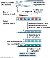
Linear DNA molecules become shorter with each round of replication because DNA polymerase cannot fully replicate the 5′ ends. This leads to progressive shortening of chromosomes in somatic cells.
Telomeres and Telomerase
Telomeres are repetitive nucleotide sequences at the ends of eukaryotic chromosomes that protect genes from erosion. The enzyme telomerase extends telomeres in germ cells, maintaining chromosome integrity for future generations. Telomere shortening is associated with aging and may protect against cancer by limiting cell divisions.
Key Point: Telomerase activity is often found in cancer cells, enabling unlimited division.
Chromosome Structure and DNA Packing
DNA and Chromatin Organization
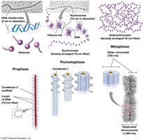
In bacteria, DNA is circular and supercoiled with minimal protein. In eukaryotes, DNA is linear and packaged with histone proteins into chromatin. The basic unit of chromatin is the nucleosome, consisting of DNA wrapped around a core of eight histones. Chromatin can be further compacted into higher-order structures, especially during cell division.
Key Point: Chromatin structure regulates gene expression and accessibility.
Euchromatin and Heterochromatin
Most interphase chromatin is loosely packed (euchromatin), allowing gene expression. Some regions, such as centromeres and telomeres, remain densely packed (heterochromatin), making gene expression difficult. Chromatin packing changes during the cell cycle and can be regulated by chemical modifications of histones.
Key Point: Chromatin condensation and modification play crucial roles in gene regulation and chromosome structure.
