 Indietro
The Molecular Basis of Inheritance: DNA Structure, Replication, and Technology
Guida di studio - Note intelligenti
 Appunti personalizzati basati sui tuoi materiali, ampliati con definizioni chiave, esempi e contesto.
Ch.13 - The Molecular Basis of Inheritance
Griffith's Experiment and Bacterial Transformation
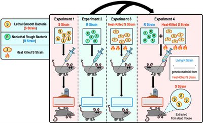
The Griffith experiment (1928) was a foundational study in molecular genetics, demonstrating that bacteria can acquire new traits through the uptake of genetic material from their environment. This process is known as transformation.
Transformation: The uptake and incorporation of external DNA by a cell, resulting in a change in genotype and phenotype.
Key Findings: Griffith showed that non-lethal rough (R) bacteria could become lethal smooth (S) bacteria when mixed with heat-killed S strain, indicating the transfer of genetic material.
Significance: This experiment suggested that a "genetic factor" (later identified as DNA) controls traits in organisms.
Example: The transformation of R strain bacteria into S strain by genetic material from heat-killed S strain.
The Hershey-Chase Experiment
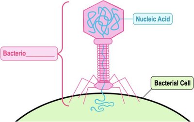
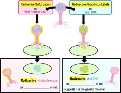
In 1952, Hershey and Chase used bacteriophages (viruses that infect bacteria) to confirm that DNA, not protein, is the genetic material.
Bacteriophage: A virus that infects bacteria, consisting of a protein coat and nucleic acid (DNA or RNA).
Experimental Design: Radioactive sulfur labeled viral protein, radioactive phosphorus labeled viral DNA.
Key Result: Only viral DNA entered the bacterial cell and directed viral replication, confirming DNA as the genetic material.
Chargaff's Rules
Erwin Chargaff discovered two key principles about DNA composition in the early 1950s:
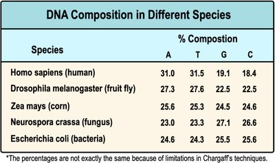
Base Composition Varies: The proportion of DNA bases (A, T, G, C) varies between species.
Base Pairing: In each species, the percentage of adenine (A) is roughly equal to thymine (T), and guanine (G) is roughly equal to cytosine (C).
Example: Human DNA contains about 31% A, 31.5% T, 19.1% G, and 18.4% C.
Species | A (%) | T (%) | G (%) | C (%) |
|---|---|---|---|---|
Homo sapiens (human) | 31.0 | 31.5 | 19.1 | 18.4 |
Drosophila melanogaster (fruit fly) | 27.3 | 27.6 | 22.5 | 22.5 |
Zea mays (corn) | 25.6 | 25.3 | 24.5 | 24.6 |
Neurospora crassa (fungus) | 23.0 | 23.3 | 27.1 | 26.6 |
Escherichia coli (bacteria) | 24.6 | 24.3 | 25.5 | 25.6 |
Discovery of DNA Structure
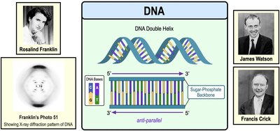
Rosalind Franklin's X-ray diffraction studies (Photo 51) and the work of James Watson and Francis Crick led to the description of DNA's double helix structure in 1953.
Double Helix: DNA consists of two antiparallel strands forming a helical structure.
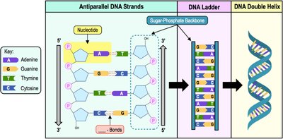
Base Pairing: Nucleotides pair via hydrogen bonds: A–T and C–G.
Antiparallel Strands: The two strands run in opposite directions (5' to 3' and 3' to 5').
Detailed DNA Structure
DNA is composed of nucleotide monomers linked together, forming two strands with distinct 5' and 3' ends.
5' End: Free phosphate group.
3' End: Free hydroxyl (-OH) group.
Phosphodiester Bond: Links the phosphate group of one nucleotide to the 3' OH of another.
Meselson-Stahl Experiment: DNA Replication Models
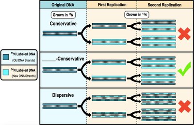
Meselson and Stahl (1958) demonstrated that DNA replication is semi-conservative, meaning each new DNA molecule contains one old (parental) strand and one newly synthesized strand.
Semi-Conservative Model: Parental strands separate and serve as templates for new strands.
Other Models: Conservative (both old strands stay together) and dispersive (mixed segments).
Introduction to DNA Replication
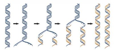
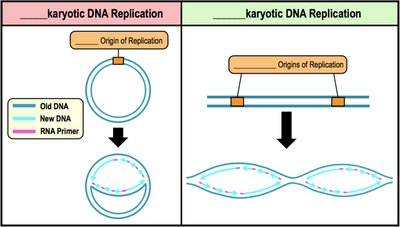
DNA replication is a fundamental process in both prokaryotes and eukaryotes, involving the separation of parental strands and synthesis of new complementary strands.
Replication Origin (ORI): Specific DNA sequence where replication begins.
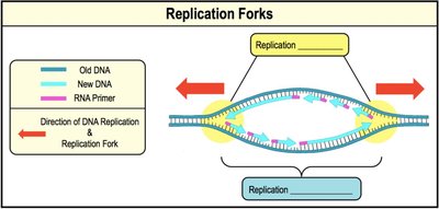
Replication Fork: Y-shaped region where DNA is unwound and replication occurs.
Bidirectional Replication: Replication proceeds in both directions from the origin.
Unwinding DNA: Topoisomerase, Helicase, and SSBs
Several proteins are involved in unwinding DNA during replication:
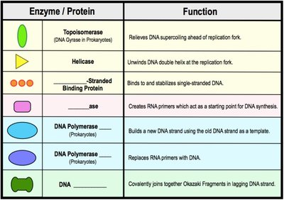
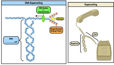
Topoisomerase: Relieves strain caused by supercoiling ahead of the replication fork.
Helicase: Unwinds the DNA double helix by breaking hydrogen bonds.
Single-Strand Binding Proteins (SSBs): Stabilize separated DNA strands and prevent reannealing.
DNA Polymerases and Their Requirements
DNA polymerases are the primary enzymes responsible for synthesizing new DNA strands.
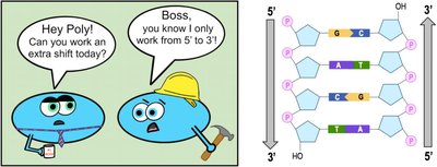
Directionality: DNA polymerases synthesize DNA in the 5' to 3' direction.
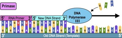
Requirements: Template strand, RNA primer (created by primase), and free nucleotides.
Types: DNA Polymerase III (elongation), DNA Polymerase I (removes RNA primers and replaces with DNA).
Leading and Lagging DNA Strands
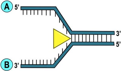
During replication, the two new DNA strands are synthesized differently due to the antiparallel nature of DNA.
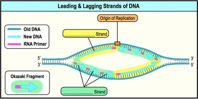
Leading Strand: Synthesized continuously in the same direction as the replication fork movement; requires only one RNA primer.
Lagging Strand: Synthesized discontinuously in the opposite direction, forming short segments called Okazaki fragments, each requiring an RNA primer.
DNA Ligase: Joins Okazaki fragments to form a continuous strand.
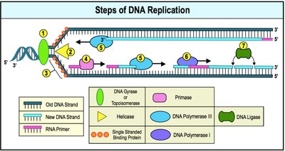
Steps of DNA Replication
DNA replication in prokaryotes can be summarized in several steps:
Topoisomerase relieves supercoiling strain.
Helicase unwinds the DNA strands.
Single-Stranded Binding Proteins stabilize separated strands.
Primase adds RNA primers.
DNA Polymerase III elongates new DNA strands.
DNA Polymerase I replaces RNA primers with DNA.
DNA Ligase joins Okazaki fragments.
 *Additional info: The above notes expand on brief points from the original material, providing definitions, examples, and context for each concept. All included images directly reinforce the explanations and are essential for visualizing the described processes.*
