 Indietro
The Plasma Membrane and Cytoskeleton: Structure, Function, and Cellular Transport
Guida di studio - Note intelligenti
 Appunti personalizzati basati sui tuoi materiali, ampliati con definizioni chiave, esempi e contesto.
The Plasma Membrane
Functions of the Plasma Membrane
The plasma membrane is a critical cellular structure that separates the internal environment of the cell from the external environment. It performs several essential functions:
Physical isolation and protection: Acts as a barrier, protecting cellular contents.
Regulation of exchange: Controls the movement of substances in and out of the cell.
Structural support: Provides shape and mechanical stability.
Identification: Contains molecules for cell recognition and signaling.
Structure of the Plasma Membrane
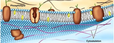
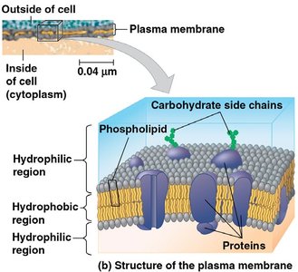
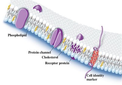
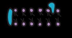
The plasma membrane is primarily composed of phospholipids, which are amphipathic molecules containing both hydrophilic (water-loving) heads and hydrophobic (water-fearing) tails. This arrangement forms a bilayer, creating a stable boundary between two aqueous compartments.
Phospholipid bilayer: The fundamental structure, with hydrophilic heads facing outward and hydrophobic tails inward.
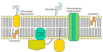
Proteins: Embedded within the bilayer, serving various functions such as transport, signaling, and structural support.
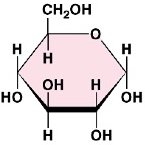
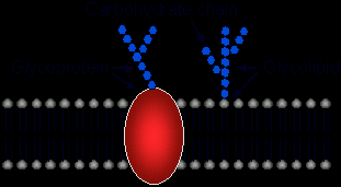
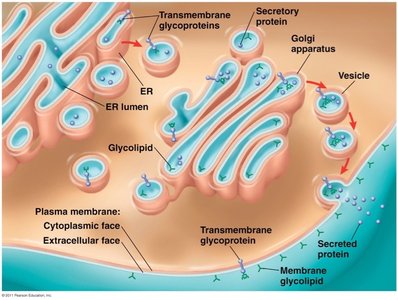
Carbohydrates: Attached to lipids (glycolipids) or proteins (glycoproteins), important for cell recognition.
Phospholipid Properties and Movements
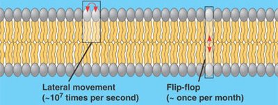
Phospholipids exhibit dynamic movements within the membrane, contributing to its fluidity:
Lateral movement: Phospholipids move side-to-side rapidly (~107 times per second).
Flip-flop movement: Rare movement of phospholipids from one leaflet to another (about once per month).
Fluid Mosaic Model
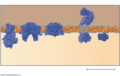
The plasma membrane is described by the fluid mosaic model, which states that the membrane is a flexible sheet of lipids with embedded proteins and attached carbohydrates. This model emphasizes the dynamic and heterogeneous nature of the membrane.
Semi-permeable: Allows selective passage of substances.
Embedded proteins: Provide specific functions such as transport and signaling.
Factors Affecting Membrane Fluidity
Membrane fluidity is influenced by several factors:
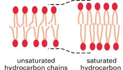
Lipid composition: Unsaturated hydrocarbon tails increase fluidity, while saturated tails decrease it.
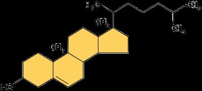
Cholesterol: Acts as a fluidity buffer, preventing tight packing at low temperatures and restricting movement at high temperatures.
Cholesterol in the Plasma Membrane
Cholesterol is a steroid and an essential structural component of mammalian membranes. It regulates membrane fluidity and permeability, with different effects depending on temperature:
Cold temperatures: Prevents tight packing of phospholipids, maintaining fluidity.
Warm temperatures: Restricts phospholipid movement, reducing fluidity.
Membrane Proteins
Membrane proteins are diverse and determine most of the membrane’s specific functions. They are classified as:
Peripheral proteins: Loosely attached to the membrane surface.
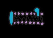
Integral proteins: Embedded within the membrane; includes transmembrane proteins that span the bilayer.

Functions of Membrane Proteins
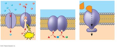
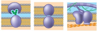
Membrane proteins perform a variety of functions:
Transport: Facilitate movement of substances across the membrane.
Enzymatic activity: Catalyze reactions at the membrane surface.
Signal transduction: Relay signals from outside to inside the cell.
Cell-cell recognition: Allow cells to identify each other.
Intercellular joining: Connect cells together.
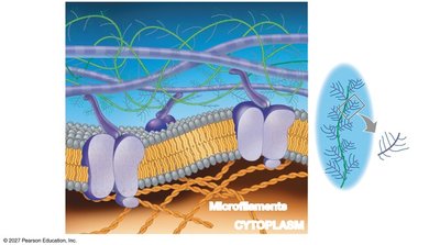
Attachment: Anchor the membrane to the cytoskeleton and extracellular matrix.
Glycolipids and Glycoproteins
Glycolipids and glycoproteins are carbohydrates covalently bonded to lipids and proteins, respectively. They play key roles in cell recognition and signaling.
Cell Walls and Extracellular Matrix
Plant Cell Walls
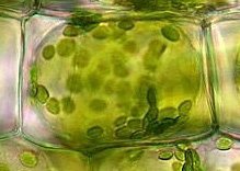
Plant cells possess a rigid cell wall that distinguishes them from animal cells. The cell wall provides protection, maintains cell shape, and is primarily composed of cellulose embedded in other carbohydrates and proteins.
Intercellular Junctions
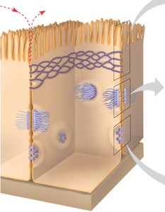
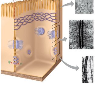
Cells in tissues adhere and communicate through specialized junctions:
Tight junctions: Prevent leakage of extracellular fluid.
Desmosomes: Anchor cells together into strong sheets.
Gap junctions: Provide cytoplasmic channels for communication.
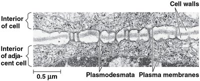
Plasmodesmata: Unique to plant cells, allow passage of water and solutes between cells.
Extracellular Matrix (ECM) in Animal Cells
Animal cells lack cell walls but are surrounded by an elaborate extracellular matrix (ECM) composed of glycoproteins (such as collagen) and other substances. The ECM holds cells together, provides structural support, and filters materials between tissues.
The Cytoskeleton
Cytoskeleton Functions
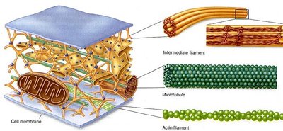
The cytoskeleton is a network of protein fibers that supports the cell, maintains its shape, anchors organelles, and enables cell movement and transport of materials.
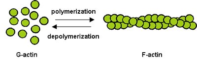
Microfilaments (actin): Enable cell movement and shape changes.
Intermediate filaments (keratin): Provide structural strength and anchor organelles.
Microtubules: Largest element, involved in cell division and rapid length changes.
Dynamic Instability of Cytoskeletal Elements
Microtubules exhibit dynamic instability, rapidly growing and shrinking, which is essential for their roles in cell division and intracellular transport.
Cilia and Flagella
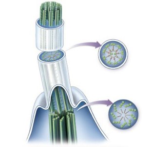
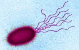
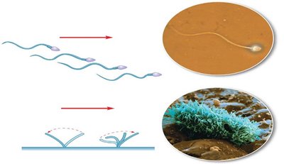
Cilia and flagella are locomotor appendages composed of microtubules. They share a similar structure (9+2 arrangement) but differ in function:
Flagella: Long appendages used for cell propulsion.
Cilia: Short appendages used for movement or moving fluid past the cell.
Motor Proteins and Cell Motility
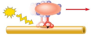
Motor proteins interact with the cytoskeleton to facilitate cell motility and transport. They use ATP to "walk" vesicles along cytoskeletal fibers.
Centrosome and Cell Division
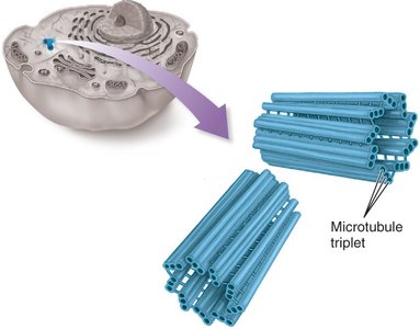
The centrosome is the microtubule-organizing center (MTOC) in animal cells, consisting of a pair of centrioles and radiating microtubules. It plays a crucial role in organizing microtubules during cell division.
Microfilaments and Microvilli
Microfilaments (actin) bear tension and resist pulling forces within the cell. They also increase membrane surface area, especially in structures like microvilli, which are important for absorption.
Actin and Cellular Motility
Actin microfilaments are involved in cellular motility, often working with the protein myosin to produce movement.
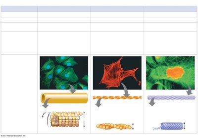
Summary Table: Cytoskeletal Elements
The following table summarizes the structure and function of the three primary cytoskeletal fibers:
Property | Microtubules | Microfilaments | Intermediate Filaments |
|---|---|---|---|
Structure | Hollow tubes | Two intertwined strands | Fibrous proteins coiled into cables |
Diameter | 25 nm | 7 nm | 8–12 nm |
Protein Subunits | Tubulin (α and β) | Actin | Keratin and others |
Main Functions | Cell division, motility, organelle movement | Cell shape, movement, muscle contraction | Structural support, anchoring organelles |
Dynamic Instability | Yes | No | No |
Key Vocabulary
Dynamic instability: Rapid growth and shrinkage of microtubules.
Semi-permeable: Selective passage of substances.
Glycolipid: Carbohydrate attached to lipid.
Glycoprotein: Carbohydrate attached to protein.
Intercellular junctions: Structures connecting cells.
Microvilli: Protrusions increasing surface area.
Study Guide Questions
What does it mean for a membrane to be semi-permeable and a fluid mosaic?
Describe the components found in all membranes. Is there a difference between the cell and plasma membrane?
What effect does cholesterol have on membrane fluidity?
What effect do fatty acids have on membrane fluidity and permeability?
Name the various types of membrane proteins and describe how they are attached to the cell membrane.
Name the various cellular functions of membrane proteins.
What are the functions of the cytoskeleton?
Name the 3 intercellular junctions found in animal cells. How do they differ from each other?
Name and describe the one junction unique to plant cells.
Name 3 functions of the extracellular matrix.
Name the 3 primary fibers of the cytoskeleton. What are they built from? Name various locations where they are found. Which have dynamic instability?
Name the similarities and differences between cilia and flagella. How do motor proteins assist them?
