 Indietro
The Structure and Function of Cellular Membranes
Guida di studio - Note intelligenti
 Appunti personalizzati basati sui tuoi materiali, ampliati con definizioni chiave, esempi e contesto.
The Membrane: Structure and Function
Overview of Membrane Composition
Cellular membranes are essential structures that define the boundaries of cells and organelles, controlling the movement of substances in and out. They are primarily composed of lipids, proteins, and carbohydrates, with phospholipids being the most abundant lipid type. The unique arrangement of these molecules allows membranes to be both stable and dynamic, supporting a variety of cellular functions.
Phospholipid Bilayer Structure
The fundamental structure of the membrane is the phospholipid bilayer. Each phospholipid molecule is amphipathic, containing a hydrophilic ("water-loving") head and two hydrophobic ("water-fearing") tails. This dual nature causes phospholipids to arrange themselves into a bilayer, with hydrophobic tails facing inward, shielded from water, and hydrophilic heads facing outward toward the aqueous environment.
Hydrophilic head: Interacts with water on both sides of the membrane.
Hydrophobic tail: Avoids water, forming the interior of the bilayer.
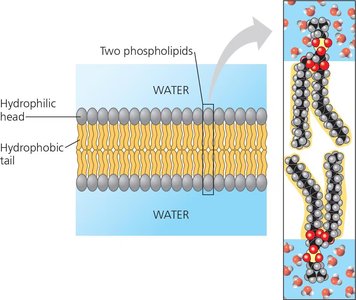
Example: The plasma membrane of all cells is a phospholipid bilayer, providing a selective barrier between the cell and its environment.
The Fluid Mosaic Model
The fluid mosaic model describes the membrane as a dynamic structure with proteins embedded or associated with a fluid lipid bilayer. Proteins and lipids can move laterally within the layer, giving the membrane flexibility and allowing for the distribution of membrane components.
Integral proteins: Span the membrane or are embedded within it.
Peripheral proteins: Loosely attached to the membrane surface.
Carbohydrates: Attached to proteins (glycoproteins) or lipids (glycolipids) on the extracellular surface, playing roles in cell recognition.
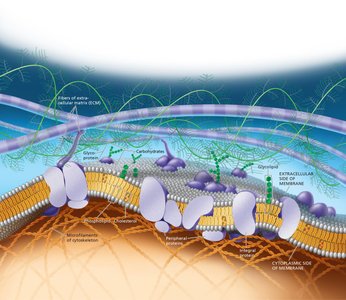
Additional info: The arrangement of proteins and lipids is not random; specialized patches called lipid rafts may exist, though their presence in living cells is debated.
Membrane Fluidity
Membranes are not rigid; their fluidity is crucial for function. Fluidity is influenced by the types of fatty acids in phospholipids and the presence of cholesterol.
Unsaturated hydrocarbon tails: Have kinks that prevent tight packing, increasing fluidity.
Saturated hydrocarbon tails: Pack closely, making the membrane more viscous.
Cholesterol: Acts as a "fluidity buffer," reducing fluidity at moderate temperatures but preventing solidification at low temperatures.
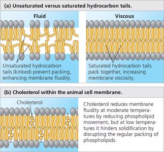
Example: Fish living in cold environments have membranes rich in unsaturated fatty acids to maintain fluidity.
Experimental Evidence for Membrane Fluidity
Experiments have demonstrated that membrane proteins can move within the plane of the membrane. For example, when mouse and human cells are fused, their membrane proteins intermingle over time, indicating lateral movement.
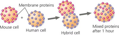
Key Point: Membrane proteins are not static; their movement is essential for many cellular processes.
Membrane Proteins: Types and Functions
Membrane proteins are diverse and perform a wide range of functions. They are classified as integral (including transmembrane) or peripheral proteins.
Integral proteins: Penetrate the hydrophobic core; many are transmembrane proteins spanning the bilayer.
Peripheral proteins: Attached to the membrane surface, often to integral proteins or lipids.
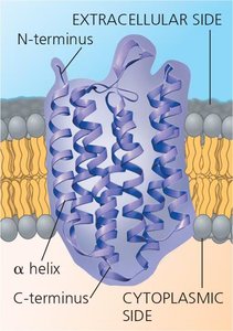
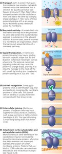
Major functions of membrane proteins include:
Transport: Moving substances across the membrane.
Enzymatic activity: Catalyzing reactions at the membrane surface.
Signal transduction: Relaying signals from outside to inside the cell.
Cell-cell recognition: Identifying other cells via surface molecules.
Intercellular joining: Connecting adjacent cells.
Attachment: Linking to the cytoskeleton and extracellular matrix for structural support.
Membrane Carbohydrates and Cell-Cell Recognition
Carbohydrates attached to the membrane's exterior surface play a critical role in cell-cell recognition. These short, branched chains are usually covalently bonded to proteins (forming glycoproteins) or lipids (forming glycolipids). Their diversity allows cells to distinguish one another, which is vital for immune response and tissue formation.
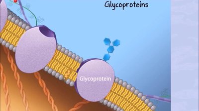
Example: Blood type antigens are determined by specific carbohydrate patterns on red blood cell membranes.
Medical Relevance: HIV and Membrane Proteins
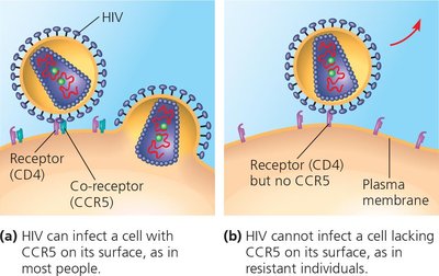
Some viruses, such as HIV, exploit membrane proteins to infect cells. HIV binds to the CD4 receptor and a co-receptor (CCR5) on the surface of immune cells. Individuals lacking CCR5 are resistant to HIV infection, highlighting the importance of membrane proteins in disease susceptibility.
Synthesis and Sidedness of Membranes
Membranes have distinct inside (cytoplasmic) and outside (extracellular) faces, with specific orientation of proteins and carbohydrates. This asymmetry is established during membrane synthesis in the endoplasmic reticulum (ER) and Golgi apparatus and is maintained as vesicles transport membrane components to the plasma membrane.
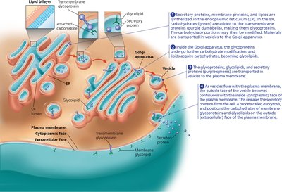
Key Point: The sidedness of membranes is crucial for their function, ensuring that specific molecules are presented on the correct side of the cell.
Summary Table: Key Components of the Plasma Membrane
Component | Structure | Function |
|---|---|---|
Phospholipids | Bilayer with hydrophilic heads and hydrophobic tails | Forms the basic structure; barrier to most water-soluble substances |
Proteins (Integral & Peripheral) | Embedded or attached to bilayer | Transport, signaling, enzymatic activity, cell recognition, joining, attachment |
Carbohydrates | Short chains attached to proteins/lipids | Cell-cell recognition, protection |
Cholesterol | Interspersed among phospholipids | Modulates fluidity and stability |
