 Indietro
Tour of the Cell: Structure, Function, and Methods of Study
Guida di studio - Note intelligenti
 Appunti personalizzati basati sui tuoi materiali, ampliati con definizioni chiave, esempi e contesto.
Cell Structure and Diversity
Cell Shapes and Sizes
Cells exhibit a wide variety of shapes and sizes, reflecting their specialized functions in multicellular organisms. The diversity in cell morphology is essential for the proper functioning of tissues and organs.
Cell Shape: Cells can be round, elongated, flat, or irregular, depending on their role.
Cell Size: Cell size ranges from microscopic bacteria to large egg cells. Most plant and animal cells are between 10–100 μm in diameter.
Surface Area to Volume Ratio: Cells are limited in size because as they grow, their volume increases faster than their surface area, affecting nutrient uptake and waste removal.
Example: Nerve cells are long and thin to transmit signals, while red blood cells are biconcave for gas exchange.
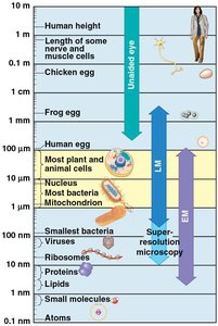
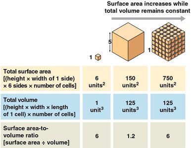
Additional info: The surface area-to-volume ratio is mathematically expressed as , and is critical for cell efficiency.
Prokaryotic Cells: Bacteria
Bacterial Cell Wall Composition and Classification
Bacteria are classified based on the structure and composition of their cell walls, which is crucial for their identification and response to antibiotics.
Gram-positive bacteria: Have thick peptidoglycan layers and lack an outer membrane.
Gram-negative bacteria: Have thin peptidoglycan layers and an additional outer membrane.
Gram Stain: A laboratory technique used to differentiate bacteria based on cell wall properties.
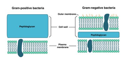
Additional info: Gram-positive bacteria stain purple, while Gram-negative bacteria stain pink due to differences in cell wall structure.
Eukaryotic Cells: Animal, Plant, and Fungus Cells
Standard Animal Cell Organelles
Animal cells contain various organelles, each with specialized functions necessary for cellular metabolism, growth, and communication.
Nucleus: Contains genetic material (DNA).
Endoplasmic Reticulum (ER): Smooth ER synthesizes lipids; Rough ER is involved in protein synthesis.
Golgi Apparatus: Modifies, sorts, and packages proteins and lipids.
Ribosomes: Sites of protein synthesis.
Mitochondria: Powerhouse of the cell, site of cellular respiration.
Cytoskeleton: Provides structural support and facilitates intracellular transport.
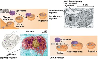
Lysosomes: Digestive organelles for waste processing.
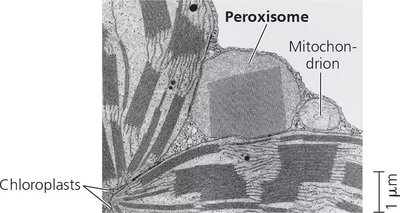
Peroxisomes: Break down fatty acids and detoxify harmful substances.
Standard Plant Cell Organelles
Plant cells possess unique organelles and structures that distinguish them from animal cells, including chloroplasts and a central vacuole.
Chloroplast: Site of photosynthesis.
Central Vacuole: Stores water, nutrients, and waste products.
Other Plastids: Storage and synthesis of pigments and starch.
Dictyosomes: Plant-specific Golgi bodies involved in cell wall formation.
Standard Fungus Cell Organelles
Fungal cells share many organelles with animal and plant cells but have unique features such as spindle pole bodies and specialized vacuoles.
Spindle Pole Bodies: Organize microtubules during cell division.
Vacuoles: Storage and waste processing.
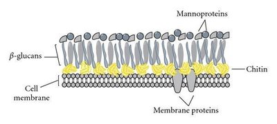
Additional info: Fungal cell walls contain chitin, β-glucans, and mannoproteins, providing rigidity and protection.
Microscopy and Cell Study Methods
Types of Microscopes
Microscopes are essential tools for studying cells and their organelles. Different types of microscopes offer varying levels of resolution and magnification.
Light Microscope (LM): Uses visible light to observe cells and tissues.
Scanning Electron Microscope (SEM): Provides detailed surface images of cells.
Transmission Electron Microscope (TEM): Reveals internal structures of cells at high resolution.
Attomicroscope: Advanced instrument for visualizing electron movement.
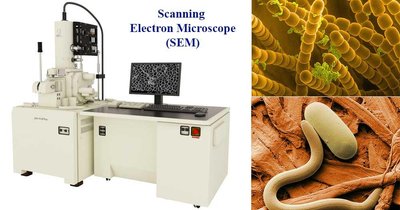
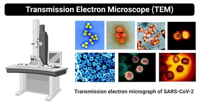
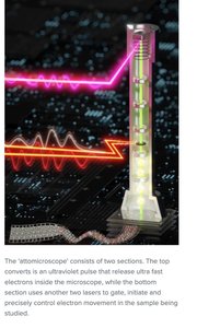
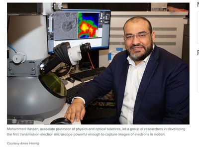
Additional info: Electron microscopes achieve much higher resolution than light microscopes, allowing visualization of subcellular structures.
Cell Fractionation
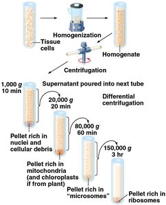
Cell fractionation is a laboratory technique used to separate cellular components based on size and density, enabling detailed study of organelles.
Homogenization: Cells are broken up to release their contents.
Centrifugation: Separates organelles by spinning at different speeds.
Differential Centrifugation: Sequentially isolates nuclei, mitochondria, microsomes, and ribosomes.
Cytoskeleton
Types of Cytoskeletal Fibers
The cytoskeleton is a network of protein fibers that provides structural support, facilitates cell movement, and organizes organelles.
Microtubules: Thickest fibers, composed of tubulin; involved in cell shape, transport, and division.
Microfilaments (Actin Filaments): Thinnest fibers; support cell shape and enable movement.
Intermediate Filaments: Provide mechanical strength; intermediate in size.
Motor Proteins: Work with the cytoskeleton to transport vesicles and organelles within cells, powered by ATP.
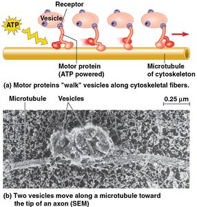
Additional info: Microtubules are essential for mitosis, while actin filaments are crucial for muscle contraction and cell motility.
Extracellular Structures and Cell Junctions
Plant Cell Wall Layers
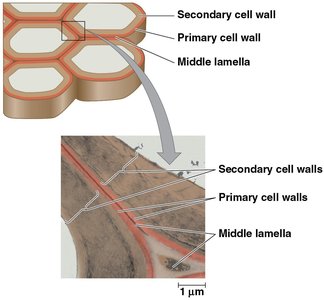
Plant cells are surrounded by a complex cell wall composed of multiple layers, each with distinct functions and compositions.
Primary Cell Wall: Thin and flexible, formed first.
Middle Lamella: Rich in pectins, glues adjacent cells together.
Secondary Cell Wall: Thicker, provides additional strength.
Dictyosomes: Vesicles involved in cell wall synthesis.
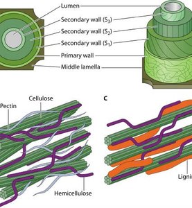
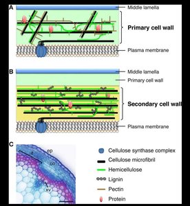
Additional info: Cellulose, hemicellulose, pectin, and lignin are key components of plant cell walls.
Extracellular Matrix (ECM) in Animal Cells
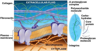
The ECM is a network of proteins and carbohydrates outside animal cells, providing structural support and facilitating cell communication.
Collagen: Main structural protein.
Proteoglycans: Protein-polysaccharide complexes.
Fibronectin: Glycoprotein involved in cell adhesion.
Cell Junctions
Cells communicate and adhere to each other through specialized junctions, which vary between plants, fungi, and animals.
Plasmodesmata (Plants): Channels for transport and communication between plant cells.
Septal Pores (Fungi): Allow communication between fungal cells.
Animal Cell Junctions:
Tight Junctions: Prevent leakage between cells.
Anchoring Junctions (Desmosomes): Provide mechanical stability.
Gap Junctions: Allow direct communication and transfer of ions and molecules.
Additional info: Cell junctions are essential for tissue integrity and coordinated cellular activity.
Summary Table: Cytoskeletal Fibers
The following table summarizes the main features of the three types of cytoskeletal fibers:
Type | Description | Size | Example | Roles |
|---|---|---|---|---|
Microtubule | Hollow tubes of tubulin | 25 nm diameter | Cilia, flagella, mitotic spindle | Cell shape, transport, division |
Intermediate Filament | Fibrous proteins coiled into cables | 8–12 nm diameter | Nuclear lamina, keratin | Mechanical support |
Microfilament | Actin filaments | 7 nm diameter | Muscle fibers, cell cortex | Cell movement, shape |
Summary Table: Cell Junctions in Animals
Junction Type | Description | Example |
|---|---|---|
Tight Junction | Seals cells together, prevents leakage | Intestinal epithelium |
Anchoring Junction (Desmosome) | Connects cytoskeletons, provides strength | Skin tissue |
Gap Junction | Allows passage of ions and molecules | Cardiac muscle |
