Indietro

Viruses and Eukaryotic Cell Structures: Study Guide for General Biology

Guida di studio - Note intelligenti

Appunti personalizzati basati sui tuoi materiali, ampliati con definizioni chiave, esempi e contesto.

Viruses

Impact of Viruses on Humans

Viruses are infectious agents that can invade diverse tissues in the human body, causing a wide range of diseases. They have played a significant role in human evolution, as viral DNA can become integrated into the human genome. In modern times, viruses are responsible for epidemics and pandemics, affecting global health and population dynamics.

  • Infectious Diversity: Viruses can target various tissues, including the nervous system, respiratory tract, digestive organs, and skin.

  • Evolutionary Influence: Some viral DNA sequences are found in the human genome, indicating past infections and evolutionary adaptation.

  • Epidemics and Pandemics: Viruses such as influenza, HIV, and coronaviruses have caused widespread outbreaks.
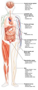
Human body showing tissues infected by different viruses

  • Population Impact: Historical pandemics, such as smallpox and influenza, have caused dramatic declines in population and life expectancy.
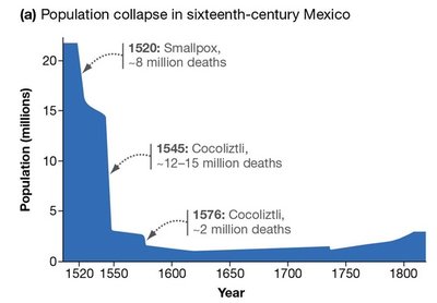
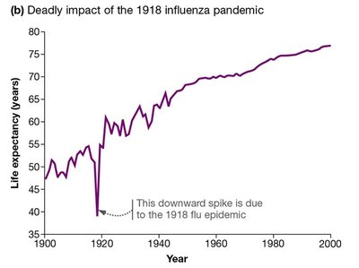
Population collapse in sixteenth-century Mexico due to viral epidemics Life expectancy drop during the 1918 influenza pandemic

Classification of Viruses

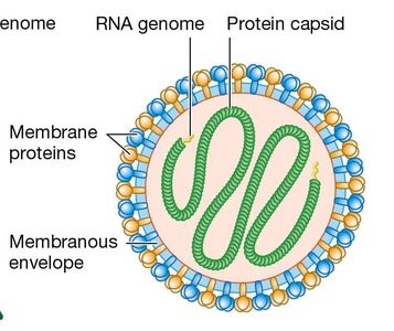
Viruses are classified based on their genomic material and structural features. The genome can be DNA or RNA, and the viral shell (capsid) can be icosahedral or enveloped.

  • Genomic Material: Viruses may contain DNA or RNA as their genetic material.

  • Capsid: The protein shell that encases the genome; can be icosahedral or helical.

  • Envelope: Some viruses have a lipid membrane surrounding the capsid, derived from the host cell.
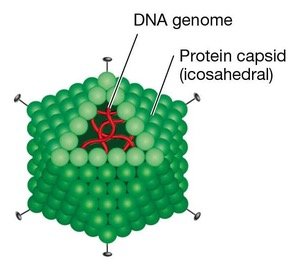
Adenovirus structure with DNA genome and protein capsid Measles virus structure with RNA genome and protein capsid

Viral Replication and Propagation

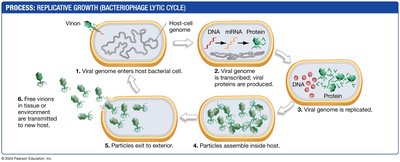
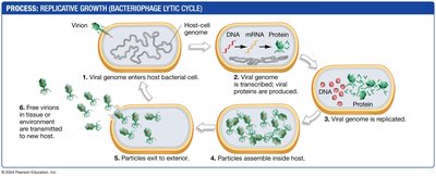
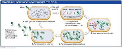
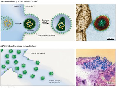
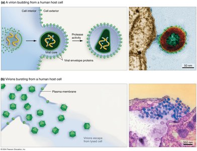
Viruses reproduce by entering host cells, hijacking cellular machinery to replicate their genome and produce new viral particles. The lytic cycle is a common mode of viral propagation, resulting in cell lysis and release of new virions.

  • Lytic Cycle: Virus enters cell, replicates genome, assembles new particles, and lyses the host cell to release virions.

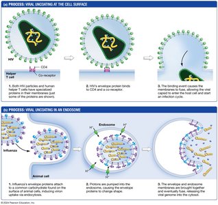
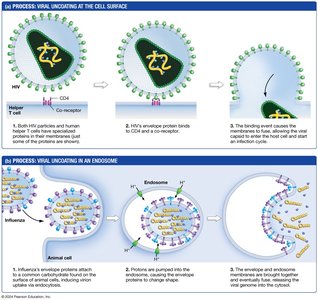
  • Enveloped Viruses: Enter cells via membrane fusion, as seen in HIV.

  • Endocytosis: Some viruses, like influenza, enter cells through endocytosis.
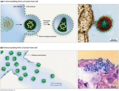
Viruses entering cells and reproducing Viral replication and propagation Lytic cycle of viral propagation Lytic cycle of viral propagation Enveloped viruses entering cells via fusion HIV virus entry via fusion HIV virus entry via fusion HIV virus entry via fusion Lytic cycle of viral propagation Lytic cycle of viral propagation Enveloped viruses entering cells via fusion HIV virus entry via fusion HIV virus entry via fusion HIV virus entry via fusion Lytic cycle of viral propagation Lytic cycle of viral propagation Enveloped viruses entering cells via fusion HIV virus entry via fusion HIV virus entry via fusion HIV virus entry via fusion Lytic cycle of viral propagation Lytic cycle of viral propagation Enveloped viruses entering cells via fusion HIV virus entry via fusion HIV virus entry via fusion HIV virus entry via fusion Lytic cycle of viral propagation Lytic cycle of viral propagation Enveloped viruses entering cells via fusion HIV virus entry via fusion HIV virus entry via fusion HIV virus entry via fusion Lytic cycle of viral propagation Lytic cycle of viral propagation Enveloped viruses entering cells via fusion HIV virus entry via fusion HIV virus entry via fusion HIV virus entry via fusion Lytic cycle of viral propagation Lytic cycle of viral propagation Enveloped viruses entering cells via fusion HIV virus entry via fusion HIV virus entry via fusion HIV virus entry via fusion Lytic cycle of viral propagation Lytic cycle of viral propagation Enveloped viruses entering cells via fusion HIV virus entry via fusion HIV virus entry via fusion HIV virus entry via fusion Lytic cycle of viral propagation Lytic cycle of viral propagation Enveloped viruses entering cells via fusion HIV virus entry via fusion HIV virus entry via fusion HIV virus entry via fusion Lytic cycle of viral propagation Lytic cycle of viral propagation Enveloped viruses entering cells via fusion HIV virus entry via fusion HIV virus entry via fusion HIV virus entry via fusion Lytic cycle of viral propagation Lytic cycle of viral propagation Enveloped viruses entering cells via fusion HIV virus entry via fusion HIV virus entry via fusion HIV virus entry via fusion Lytic cycle of viral propagation Lytic cycle of viral propagation Enveloped viruses entering cells via fusion HIV virus entry via fusion HIV virus entry via fusion HIV virus entry via fusion Lytic cycle of viral propagation Lytic cycle of viral propagation Enveloped viruses entering cells via fusion HIV virus entry via fusion HIV virus entry via fusion HIV virus entry via fusion Lytic cycle of viral propagation Lytic cycle of viral propagation Enveloped viruses entering cells via fusion HIV virus entry via fusion HIV virus entry via fusion HIV virus entry via fusion Lytic cycle of viral propagation Lytic cycle of viral propagation Enveloped viruses entering cells via fusion HIV virus entry via fusion HIV virus entry via fusion HIV virus entry via fusion Lytic cycle of viral propagation Lytic cycle of viral propagation Enveloped viruses entering cells via fusion HIV virus entry via fusion HIV virus entry via fusion HIV virus entry via fusion Lytic cycle of viral propagation Lytic cycle of viral propagation Enveloped viruses entering cells via fusion HIV virus entry via fusion HIV virus entry via fusion HIV virus entry via fusion Lytic cycle of viral propagation Lytic cycle of viral propagation Enveloped viruses entering cells via fusion HIV virus entry via fusion HIV virus entry via fusion HIV virus entry via fusion Lytic cycle of viral propagation Lytic cycle of viral propagation Enveloped viruses entering cells via fusion HIV virus entry via fusion HIV virus entry via fusion HIV virus entry via fusion Lytic cycle of viral propagation Lytic cycle of viral propagation Enveloped viruses entering cells via fusion HIV virus entry via fusion HIV virus entry via fusion HIV virus entry via fusion Lytic cycle of viral propagation Lytic cycle of viral propagation Enveloped viruses entering cells via fusion HIV virus entry via fusion HIV virus entry via fusion HIV virus entry via fusion Lytic cycle of viral propagation Lytic cycle of viral propagation Enveloped viruses entering cells via fusion HIV virus entry via fusion HIV virus entry via fusion HIV virus entry via fusion Lytic cycle of viral propagation Lytic cycle of viral propagation Enveloped viruses entering cells via fusion HIV virus entry via fusion HIV virus entry via fusion HIV virus entry via fusion Lytic cycle of viral propagation Lytic cycle of viral propagation Enveloped viruses entering cells via fusion HIV virus entry via fusion HIV virus entry via fusion HIV virus entry via fusion Lytic cycle of viral propagation Lytic cycle of viral propagation Enveloped viruses entering cells via fusion HIV virus entry via fusion HIV virus entry via fusion HIV virus entry via fusion Lytic cycle of viral propagation Lytic cycle of viral propagation Enveloped viruses entering cells via fusion HIV virus entry via fusion HIV virus entry via fusion HIV virus entry via fusion Lytic cycle of viral propagation Lytic cycle of viral propagation Enveloped viruses entering cells via fusion HIV virus entry via fusion HIV virus entry via fusion HIV virus entry via fusion Lytic cycle of viral propagation Lytic cycle of viral propagation Enveloped viruses entering cells via fusion HIV virus entry via fusion HIV virus entry via fusion HIV virus entry via fusion Lytic cycle of viral propagation Lytic cycle of viral propagation Enveloped viruses entering cells via fusion HIV virus entry via fusion HIV virus entry via fusion HIV virus entry via fusion

Eukaryotic Cell Structures

Overview of Eukaryotic Cells

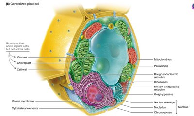
Eukaryotic cells are complex, containing membrane-bound organelles that perform specialized functions. Animal and plant cells share many organelles, but also have unique structures.

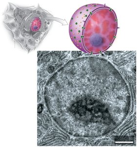
  • Nucleus: Contains genetic material and controls cellular activities.

  • Ribosomes: Sites of protein synthesis; can be free in cytoplasm or attached to rough ER.

  • Endoplasmic Reticulum (ER): Rough ER synthesizes proteins; smooth ER synthesizes lipids and processes toxins.

  • Golgi Apparatus: Modifies, sorts, and packages proteins and lipids for transport.

  • Lysosomes: Digestive organelles in animal cells, breaking down waste and old organelles.

  • Vacuoles: Large storage organelles in plant, fungal, and some protist cells.

  • Chloroplasts: Site of photosynthesis in plants and algae.

  • Mitochondria: Powerhouse of the cell, generating ATP through cellular respiration.

  • Peroxisomes: Break down fatty acids and detoxify harmful substances.
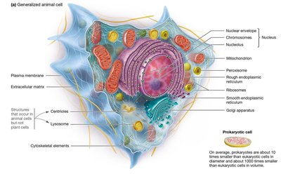
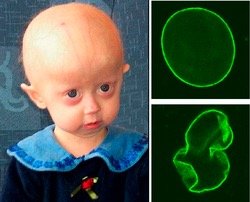
Overview of eukaryotic cells Generalized animal cell structure Generalized plant cell structure Electron microscope image of nucleus Progeria and nuclear envelope defects

Ribosomes and Endoplasmic Reticulum

Ribosomes are essential for translating mRNA into proteins. The rough ER is studded with ribosomes, while the smooth ER is involved in lipid synthesis and detoxification.

  • Free Ribosomes: Synthesize proteins for use within the cell.

  • Rough ER: Synthesizes proteins for secretion or membrane insertion.

  • Smooth ER: Synthesizes lipids and processes toxins.

Ribosomes attached to rough ER Rough ER and free ribosomes Smooth ER lumen

Lysosomes, Peroxisomes, and Vacuoles

Lysosomes are specialized for digestion and recycling in animal cells. Peroxisomes break down fatty acids and detoxify. Vacuoles serve as storage and maintain cell structure in plants, fungi, and protists.

  • Lysosomes: Contain hydrolytic enzymes for digestion.

  • Peroxisomes: Enzyme core for oxidation reactions.

  • Vacuoles: Store water, nutrients, and waste; maintain turgor pressure in plants.

Peroxisome enzyme core Material being digested within lysosomes Delivery to lysosomes for breakdown and recycling Vacuole in plant cell Vacuole in plant cell

Chloroplasts and Mitochondria

Chloroplasts and mitochondria are energy-converting organelles. Chloroplasts perform photosynthesis, while mitochondria generate ATP. Both are believed to have evolved via endosymbiosis.

  • Chloroplasts: Contain chlorophyll; convert light energy to chemical energy.

  • Mitochondria: Site of cellular respiration; contain their own DNA.

  • Endosymbiosis Theory: Suggests mitochondria and chloroplasts originated from engulfed prokaryotes.

Chloroplast structure Mitochondria structure Evolution of mitochondria via endosymbiosis Evolution of chloroplasts via endosymbiosis

Specialization and Nuclear Transport

Cells may contain varying amounts of organelles depending on their function. The nucleus is a hub of genetic activity, with constant transport of molecules through nuclear pores. Nuclear localization signals (NLS) direct proteins to the nucleus.

  • Specialized Functions: Cells adapt organelle abundance to their roles.

  • Nuclear Transport: Proteins and RNA move in and out of the nucleus via nuclear pores.

  • Nuclear Localization Signal (NLS): A sequence of amino acids (~17 AA) that targets proteins to the nucleus.

Specialized cell functions and organelle abundance Specialized cell functions and organelle abundance Specialized cell functions and organelle abundance Specialized cell functions and organelle abundance Nuclear transport Nuclear transport Nucleoplasmin nuclear signal experiment Nucleoplasmin nuclear signal experiment Nuclear localization signal experiment

Additional info:

  • Viruses are covered in General Biology Chapter 33.

  • Eukaryotic cell structure is covered in Chapter 7.

  • Endosymbiosis theory is covered in Chapter 27.

Pearson Logo

Study Prep