 Indietro
A Preview of Cell Biology: Foundations, Techniques, and Model Systems
Guida di studio - Note intelligenti
 Appunti personalizzati basati sui tuoi materiali, ampliati con definizioni chiave, esempi e contesto.
The Cell Theory: Historical Foundations
Early Microscopy and Discovery of Cells
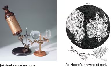
The origins of cell biology trace back over 300 years, beginning with the invention and use of early microscopes by European scientists. Robert Hooke, in 1665, constructed a microscope and observed cork, identifying boxlike compartments he termed cells (from Latin cellula, meaning "little room"). Although Hooke saw only the cell walls of dead plant tissue, his observations laid the groundwork for future studies.
Robert Hooke: First to describe cells in cork.
Antonie van Leeuwenhoek: Improved lenses, observed living cells (blood, sperm, bacteria, protozoa).
Microscope limitations: Early microscopes had low magnification and resolution, restricting detailed study.
The Cell Theory
Advances in microscopy led to the formulation of the cell theory, which is fundamental to biology. Key contributors included Robert Brown (discovered the nucleus), Matthias Schleiden (all plant tissues are composed of cells), and Theodor Schwann (animal tissues are also cellular). Rudolf Virchow later added the principle that cells arise from preexisting cells.
Principle 1: All organisms consist of one or more cells.
Principle 2: The cell is the basic unit of structure for all organisms.
Principle 3: All cells arise only from preexisting cells (omnis cellula e cellula).
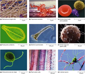
Cells exhibit remarkable diversity in shape, size, and function, reflecting their roles in different organisms and tissues.
The Emergence of Modern Cell Biology
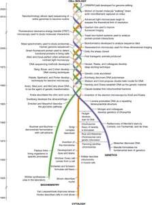
Three Strands: Cytology, Biochemistry, Genetics
Modern cell biology integrates three major disciplines: cytology (cell structure), biochemistry (cellular chemistry), and genetics (information flow). Each strand has unique historical origins and contributions, and their merging has advanced our understanding of cells.
Cytology: Study of cell structure, heavily reliant on microscopy.
Biochemistry: Study of chemical processes and molecules in cells.
Genetics: Study of inheritance and information flow.
The Cytological Strand: Cellular Structure and Microscopy
Cellular Dimensions
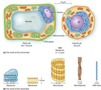
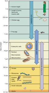
Cells and their organelles are typically too small to be seen unaided. Their sizes are measured in micrometers (μm) and nanometers (nm). Bacterial cells are a few micrometers in diameter, while plant and animal cells are larger. Organelles like mitochondria and chloroplasts are a few micrometers, and structures like ribosomes and DNA are measured in nanometers.
Micrometer (μm): 1 millionth of a meter; used for cells and organelles.
Nanometer (nm): 1 billionth of a meter; used for molecules and subcellular structures.
Angstrom (Å): 0.1 nm; used for atomic-scale measurements.
Microscopy Techniques
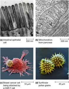
Microscopy is essential for visualizing cells and their components. The two main types are light microscopy and electron microscopy, each with distinct capabilities.
Light Microscopy: Uses visible light; includes brightfield, phase-contrast, differential interference contrast, fluorescence, and confocal microscopy.
Electron Microscopy: Uses electron beams; includes transmission (TEM) and scanning (SEM) electron microscopes, offering much higher resolution.
Resolution: The ability to distinguish two close objects as separate; electron microscopes have much greater resolving power than light microscopes.
The Biochemical Strand: Chemistry of Cells
Biochemical Reactions and Pathways
Biochemistry explores the chemical processes within cells. Key discoveries include the synthesis of organic compounds from inorganic precursors, the identification of enzymes as biological catalysts, and the elucidation of metabolic pathways such as glycolysis and the Krebs cycle.
Enzymes: Biological catalysts that accelerate chemical reactions.
ATP: Adenosine triphosphate, the principal energy storage molecule in cells.
Radioisotopes: Used to trace metabolic pathways (e.g., Calvin cycle).
Biochemistry Methods
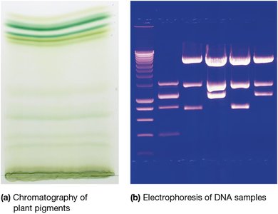
Techniques for isolating and analyzing cellular components include centrifugation, chromatography, and electrophoresis.
Centrifugation: Separates cell components by size, shape, and density.
Chromatography: Separates molecules based on size, charge, or affinity.
Electrophoresis: Uses electric fields to separate DNA, RNA, and proteins by size and charge.
Mass Spectrometry: Determines the size and composition of proteins.
The Genetic Strand: Information Flow in Cells
Classical and Molecular Genetics
Genetics investigates how traits are inherited and how genetic information is stored and expressed. Key milestones include Mendel's principles, the discovery of chromosomes, and the identification of DNA as the genetic material.
Chromosome Theory: Genes are located on chromosomes in the nucleus.
DNA: The molecule responsible for genetic inheritance.
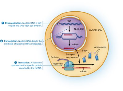
Central Dogma: Information flows from DNA to RNA to protein.
Modern Genetic Techniques
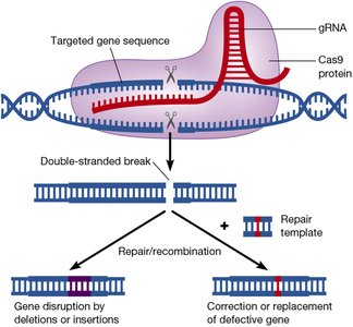
Advances in molecular genetics include recombinant DNA technology, DNA sequencing, and genome editing. The CRISPR/Cas9 system allows precise genome modifications, revolutionizing genetic research.
Restriction Enzymes: Cleave DNA at specific sequences.
DNA Cloning: Produces multiple copies of DNA sequences.
CRISPR/Cas9: Genome editing tool using guide RNA and Cas9 protein to create targeted DNA breaks.
Bioinformatics and "-Omics"
Bioinformatics merges computer science and biology to analyze large-scale genetic and protein data. Genomics, proteomics, transcriptomics, metabolomics, lipidomics, and ionomics are fields that study all genes, proteins, transcripts, metabolites, lipids, and ions in a cell, respectively.
Genomics: Study of all genes in an organism.
Proteomics: Study of all proteins in a cell.
Transcriptomics: Study of all RNA transcripts.
Metabolomics: Study of all metabolic reactions.
Scientific Inquiry and Model Systems
Experimental Design and Hypothesis Testing
Scientific knowledge in cell biology is built through hypothesis-driven experiments. Well-designed experiments alter only one variable at a time, using controls and model systems to ensure reliable results.
Hypothesis: Tentative explanation tested by experiments.
Null Hypothesis: Opposite of the hypothesis, tested for rejection.
In vitro: Experiments in glass (test tube).
In vivo: Experiments in living organisms.
In silico: Experiments using computer simulations.
Model Organisms in Cell Biology
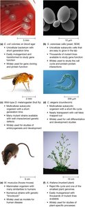
Model organisms are species that are easy to study, manipulate, and have well-characterized genetics. They are essential for understanding cellular processes and gene function.
Escherichia coli: Bacterial model for DNA replication and protein synthesis.
Saccharomyces cerevisiae: Yeast model for eukaryotic cell processes.
Drosophila melanogaster: Fruit fly model for genetics and development.
Caenorhabditis elegans: Roundworm model for cell differentiation and development.
Mus musculus: Mouse model for mammalian biology and disease.
Arabidopsis thaliana: Plant model for genetics and photosynthesis.
Summary Table: Main Microscopy Techniques
Technique | Principle | Resolution | Applications |
|---|---|---|---|
Brightfield Light Microscopy | White light passes through specimen | ~200 nm | General cell structure |
Phase-Contrast/DIC Microscopy | Enhances contrast in living cells | ~200 nm | Live cell imaging |
Fluorescence Microscopy | Detects fluorescent molecules | ~200 nm | Protein/DNA localization |
Confocal Microscopy | Laser scans single plane | ~200 nm | Thick specimen imaging |
Transmission Electron Microscopy (TEM) | Electrons transmitted through specimen | ~2 nm | Ultrastructure, organelles |
Scanning Electron Microscopy (SEM) | Electrons scan specimen surface | ~10 nm | Surface topology |
Additional info: Academic context was added to clarify the historical development, technical details, and applications of cell biology concepts and methods. The notes are structured to provide a comprehensive overview suitable for exam preparation and foundational understanding.
