 Indietro
Cell Junctions and Extracellular Structures: Structure, Function, and Clinical Relevance
Guida di studio - Note intelligenti
 Appunti personalizzati basati sui tuoi materiali, ampliati con definizioni chiave, esempi e contesto.
Cell Junctions and Extracellular Structures
Introduction to Cell Junctions and the Extracellular Matrix
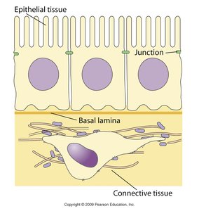
Cell junctions and the extracellular matrix (ECM) are essential for the structural integrity, communication, and function of tissues in multicellular organisms. These structures mediate cell-cell and cell-matrix interactions, contributing to tissue formation, maintenance, and signaling.
Cell junctions are specialized structures that connect adjacent cells or cells to the ECM.
Extracellular matrix (ECM) is a complex network of proteins and polysaccharides that provides structural and biochemical support to surrounding cells.
These components are critical for tissue architecture, cellular communication, and response to environmental cues.
Types of Cell-Cell Adhesion Molecules
Classification and Mechanisms
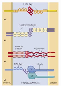
Cell adhesion molecules (CAMs) are proteins located on the cell surface involved in binding with other cells or with the ECM. They are classified based on their interaction types and molecular families.
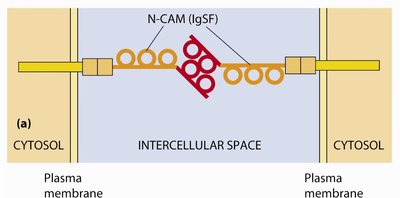
Homotypic (homophilic) interactions: CAMs bind to identical molecules on adjacent cells (e.g., cadherins, NCAMs).
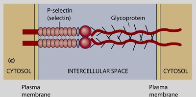
Heterotypic (heterophilic) interactions: CAMs bind to different molecules on adjacent cells (e.g., selectins binding to glycoproteins).
Main families include cadherins, immunoglobulin superfamily CAMs (IgSF), selectins, and integrins.
Immunoglobulin Superfamily CAMs (IgSF)
NCAM (Neuronal Cell Adhesion Molecule): Involved in neuronal development and growth; mutations can lead to neurological disorders.
Cadherins
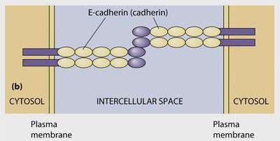
Cadherins are calcium-dependent adhesion molecules crucial for tissue formation and maintenance.
E-cadherin: Found in epithelial tissues; essential for embryonic development and tissue integrity.
N-cadherin: Important in synaptic connections in the nervous system.
Cadherin-mediated adhesion requires Ca2+ ions.
Selectins
Selectins are carbohydrate-binding proteins (lectins) that mediate transient cell-cell adhesion in the bloodstream.
P-selectin: Expressed on platelets and endothelial cells; mediates leukocyte rolling during inflammation.
L-selectin: Found on leukocytes; involved in immune cell trafficking.
Glycosylation of selectins is critical for their function.
Major Types of Cell Junctions
Overview and Classification
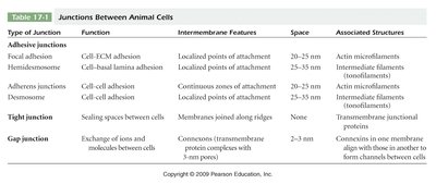
Animal cells are connected by several types of junctions, each with distinct structures and functions. These include adhesive junctions, tight junctions, and gap junctions.
Type of Junction | Function | Intermembrane Features | Space | Associated Structures |
|---|---|---|---|---|
Focal adhesion | Cell-ECM adhesion | Localized points of attachment | 20–35 nm | Actin microfilaments |
Hemidesmosome | Cell-basal lamina adhesion | Localized points of attachment | 25–35 nm | Intermediate filaments (tonofilaments) |
Adherens junction | Cell-cell adhesion | Continuous zones of attachment | 20–35 nm | Actin microfilaments |
Desmosome | Cell-cell adhesion | Localized points of attachment | 25–35 nm | Intermediate filaments (tonofilaments) |
Tight junction | Sealing spaces between cells | Membranes joined along ridges | 0 nm | Transmembrane junctional proteins |
Gap junction | Exchange of ions and molecules between cells | Connexons (transmembrane protein complexes with 3-nm pores) | 2–3 nm | Connexons in one membrane align with those in another to form channels |
Adhesive Junctions: Adherens Junctions and Desmosomes
Adhesive junctions provide strong mechanical attachments between adjacent cells or between cells and the ECM.
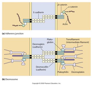
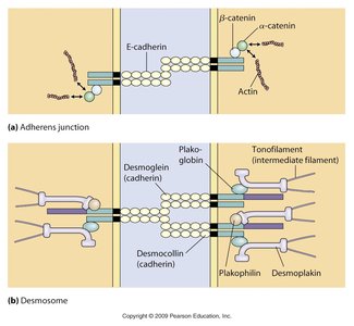
Adherens junctions: Connect actin filaments of neighboring cells via cadherins and catenins; important for maintaining tissue architecture.
Desmosomes: Connect intermediate filaments (tonofilaments) of adjacent cells via desmoglein and desmocollin; provide strong adhesion, especially in tissues under mechanical stress.
Tight Junctions
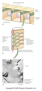
Tight junctions seal the space between epithelial cells, preventing the passage of molecules and ions through the space between cells. They are composed mainly of claudins and occludins.
Maintain cell polarity by separating the apical and basolateral surfaces of epithelial cells.
Regulate paracellular transport (movement of substances between cells).
Clinical Relevance of Cell Junctions
Genetic Diseases Affecting Cell Junctions
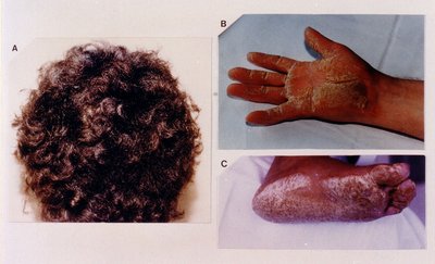
Naxos disease: Caused by mutations in plakoglobin and desmoplakin genes; leads to cardiomyopathies and skin abnormalities (e.g., woolly hair, palmoplantar keratoderma).
Extracellular Matrix (ECM) Components
Major ECM Proteins and Their Functions
The ECM is composed of a variety of proteins and polysaccharides that provide structural and functional support to cells.
Collagen: Provides tensile strength to tissues; defects can cause disorders such as Ehlers-Danlos syndrome.
Elastin: Provides elasticity; loss of elastin leads to reduced tissue flexibility (e.g., aging, Marfan syndrome).
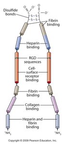
Fibronectin: Involved in cell shape, movement, and blood clotting; binds to fibrin and integrins.
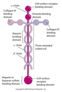
Laminin: Major component of the basal lamina; essential for cell attachment and tissue organization.
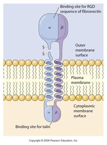
Integrins: Transmembrane receptors that connect the ECM to the cytoskeleton and mediate signal transduction.

Fibronectin and Laminin
Fibronectin: Soluble in blood, binds fibrin, and facilitates platelet attachment; critical for tissue development and wound healing.
Laminin: Present in the basal lamina; defects can cause muscular dystrophy and organ dysfunction.
Integrins and Focal Adhesions
Integrins are heterodimeric proteins that mediate cell-ECM adhesion and signal transduction. Focal adhesions are sites where integrins connect the ECM to the actin cytoskeleton, regulating cell movement and growth.
Integrins participate in "inside-out" and "outside-in" signaling, affecting cell behavior and survival.
Anchorage-dependent growth is regulated by integrin-mediated adhesion.
Collagen and Elastin: Structure and Pathology
Collagen: Triple-helical structure; crosslinking increases with age, leading to reduced flexibility.
Ehlers-Danlos syndrome: Caused by defects in collagen types I and III; leads to hyperextensible skin and joint hypermobility.
Elastin: Provides elasticity to tissues; loss with age or in Marfan syndrome leads to tissue fragility and hypermobility.
Summary Table: Major Cell Junctions and ECM Components
Structure | Main Components | Function | Clinical Relevance |
|---|---|---|---|
Adherens junction | Cadherins, catenins, actin | Cell-cell adhesion, tissue integrity | Loss in cancer metastasis |
Desmosome | Desmoglein, desmocollin, intermediate filaments | Strong cell-cell adhesion | Naxos disease, skin disorders |
Tight junction | Claudins, occludins | Barrier to paracellular transport | Leaky epithelia in disease |
Gap junction | Connexins | Cell-cell communication | Arrhythmias, deafness |
Collagen | Triple helix protein | Tensile strength | Ehlers-Danlos syndrome |
Elastin | Elastic fibers | Tissue elasticity | Marfan syndrome |
Fibronectin | Glycoprotein | Cell adhesion, migration | Wound healing defects |
Laminin | Basal lamina protein | Cell attachment | Muscular dystrophy |
Integrin | Transmembrane receptor | ECM-cytoskeleton linkage | Defective cell migration |
Additional info: This guide integrates foundational knowledge of cell junctions and ECM with clinical correlations, providing a comprehensive overview for cell biology students.
