 Indietro
Cell Membrane Structure and Function: Mini-Textbook Study Notes
Guida di studio - Note intelligenti
 Appunti personalizzati basati sui tuoi materiali, ampliati con definizioni chiave, esempi e contesto.
Membrane Structure and Function
Overview of the Plasma Membrane
The plasma membrane is a dynamic boundary that separates the internal environment of the cell from the external environment. It is essential for compartmentalization, regulation, communication, and cell joining. - Compartmentalization: Provides a distinct internal environment. - Regulation: Controls the passage of substances in and out of the cell. - Cell Communication: Facilitates signaling between cells. - Cell Joining: Enables cells to adhere to each other and form tissues. - Physical Properties: The membrane is about 10 nm thick, flexible, repairable, and expandable. 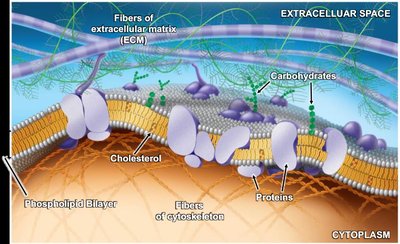
Chemical Components of the Membrane
The plasma membrane is composed of phospholipids, proteins, carbohydrates, and cholesterol. - Phospholipids: Form the basic structure of the membrane. - Proteins: Embedded or associated with the membrane, performing various functions. - Carbohydrates: Covalently bonded to proteins (glycoproteins) or lipids (glycolipids), important for cell recognition. - Cholesterol: Modulates membrane fluidity and stability.
Phospholipid Structure and Organization
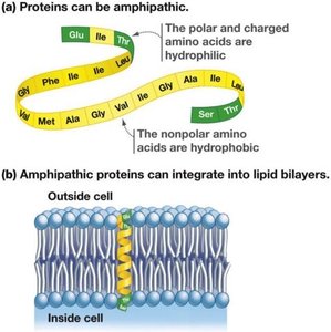
Phospholipids are amphipathic molecules with a hydrophilic (polar) head and hydrophobic (nonpolar) tails. In water, they self-assemble into higher-order structures: - Lipid Micelles: Spherical structures with hydrophilic heads facing water and hydrophobic tails inward. - Lipid Bilayers: Two layers of phospholipids with hydrophilic heads facing outward and tails facing inward, forming the basis of cell membranes. 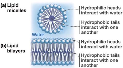 - Vesicles: Bilayers can form water-filled compartments (liposomes) in water.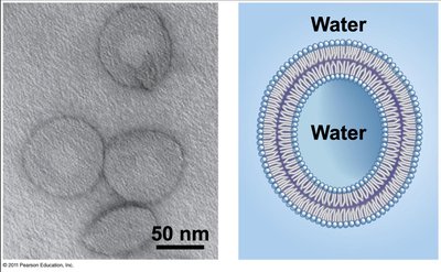
Phospholipid Bilayer Dynamics
The fluid mosaic model describes the membrane as a mosaic of proteins and other molecules embedded in a fluid phospholipid bilayer. - Fluidity: Phospholipids move laterally, rotate, and rarely flip between layers. - Permeability: Fluidity affects how easily substances cross the membrane. 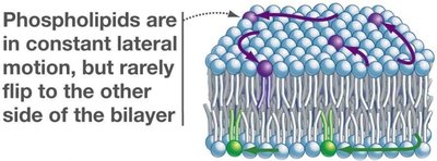
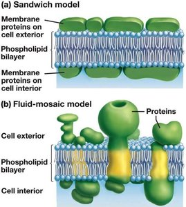
Membrane Proteins
Membrane proteins are crucial for transport, communication, enzymatic activity, and attachment. They are classified by their association with the membrane: - Integral Proteins: Have hydrophobic domains embedded in the membrane; transmembrane proteins span the entire bilayer. - Peripheral Proteins: Associate with one surface of the membrane, often interacting with integral proteins. 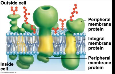
Functions of Membrane Proteins
Transport Proteins
- Transporters: Move chemicals across the membrane, including ions and large polar molecules. 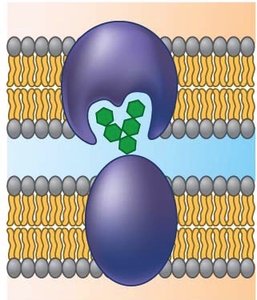
Cell Communication Proteins
- Cell Identity Markers: Glycoproteins serve as markers for cell recognition. - Receptors: Receive signals and relay messages inside the cell. 

Enzymes
- Enzymatic Proteins: Catalyze reactions in metabolic pathways. 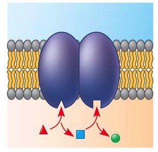


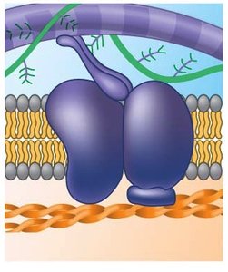
Attachment Proteins
- Attachment: Connect cells to each other (junctions) or to the extracellular matrix (ECM). 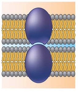
Membrane Permeability and Transport
Selective Permeability
The plasma membrane is selectively permeable, allowing some substances to cross while restricting others. - Phospholipid Bilayer: Permits small, nonpolar molecules to pass easily. - Transport Proteins: Facilitate movement of larger or polar molecules. 
Factors Affecting Membrane Fluidity
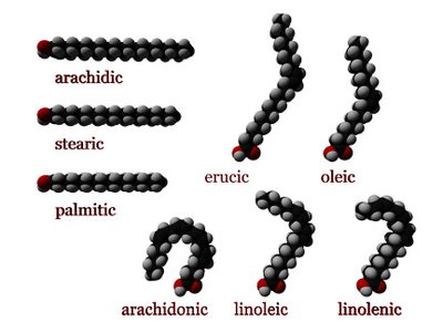
Membrane fluidity is influenced by temperature, fatty acid composition, and cholesterol content. - Temperature: Higher temperatures increase fluidity; lower temperatures decrease fluidity. 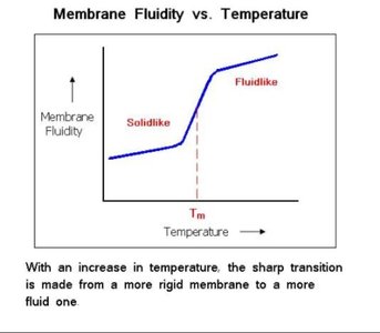 - Fatty Acid Saturation: Unsaturated fatty acids (with double bonds) create kinks, increasing fluidity and permeability. Saturated fatty acids promote tighter packing and decrease fluidity.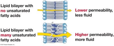
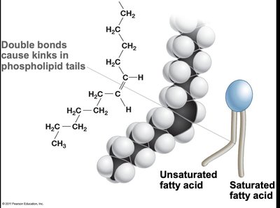 - Chain Length: Longer fatty acid tails decrease fluidity due to stronger hydrophobic interactions.
Role of Cholesterol
Cholesterol acts as a fluidity buffer, stabilizing the membrane across temperature changes. - At high temperatures: Decreases fluidity, preventing membrane disintegration. - At low temperatures: Increases fluidity, preventing membrane solidification.
Membrane Asymmetry and the Endomembrane System
Membrane Leaflets
The plasma membrane consists of two leaflets: cytoplasmic (facing the cytosol) and extracellular (facing outside). - Asymmetry: Each leaflet has distinct lipid and protein composition, established during membrane assembly in the ER and Golgi.
Extracellular Matrix and Cell Junctions
Extracellular Matrix (ECM)
Most animal cells produce and secrete a fibrous network called the ECM, which provides external support and regulates cell behavior. - Components: Collagen, elastin, polysaccharides, proteoglycans, glycoproteins. - Functions: Support, protection, cell adhesion, signaling, and tissue formation.
Cell Junctions
Cells connect to each other via junctions formed by membrane proteins, often linked to the cytoskeleton. - Tight Junctions: Prevent leakage of extracellular fluid. - Desmosomes: Fasten cells together, resist mechanical stress. - Gap Junctions: Allow exchange of ions and small molecules for cell coordination.
Mechanisms of Membrane Transport
Types of Transport
Type | Transport via | Energy Required? | Types of Substances |
|---|---|---|---|
Simple Diffusion | Lipid bilayer | No | Small and/or hydrophobic |
Facilitated Diffusion | Transporters | No | Large and/or hydrophilic |
Osmosis | Lipid bilayer/transporters | No | Water |
Active Transport | Transporters | Yes | Large and/or hydrophilic |
Bulk Transport | Vesicles | Yes | Very large substances |
Diffusion and Osmosis
- Diffusion: Net movement of molecules from high to low concentration, driven by concentration gradients. - Osmosis: Diffusion of water across a semi-permeable membrane, driven by osmotic gradients.
Facilitated Diffusion
- Channels: Tunnel-like proteins that allow specific ions or small molecules to pass. - Carriers: Proteins that bind and transport larger, hydrophilic molecules by changing shape. 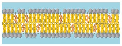
Active Transport
- Primary Active Transport: Uses ATP to move substances against their concentration gradient (e.g., Na+/K+ pump, H+ pump). - Electrogenic Pumps: Create electrochemical gradients, storing potential energy for cellular work.
Secondary Active Transport
- Uses energy stored in electrochemical gradients to move substances against their concentration gradient.
Bulk Transport
- Exocytosis: Export of large molecules via vesicles. - Endocytosis: Import of substances via vesicle formation; includes receptor-mediated endocytosis, pinocytosis, and phagocytosis.
Osmolarity, Tonicity, and Osmoregulation
Osmolarity and Osmotic Pressure
- Osmolarity: Total solute particles per liter; accounts for dissociation of ionic compounds. - Effective Osmolarity: Osmolarity of non-penetrating solutes that create osmotic gradients. - Osmotic Pressure: Pressure required to stop net water movement across a membrane.
Tonicity
- Tonicity: Ability of a solution to cause water movement by osmosis, affecting cell volume.
Effective Osmolarity Compared to Cell | Net Movement of Water | Effect on Animal Cell |
|---|---|---|
Lower | Into the cell | Swells and bursts |
Equal | None | No effect |
Higher | Out of the cell | Shrinks |
Osmoregulation
- Osmoregulation: Homeostatic processes that regulate osmolarity and water balance in cells and body fluids. - Examples: Contractile vacuole in protists, kidney function in animals, turgor pressure in plants. Additional info: These notes expand on brief points with academic context, definitions, and examples to ensure completeness and clarity for exam preparation.
