 Indietro
Cellular Membranes: Structure, Function, and Identity (Chapter 15 Study Guide)
Guida di studio - Note intelligenti
 Appunti personalizzati basati sui tuoi materiali, ampliati con definizioni chiave, esempi e contesto.
Cellular Membranes: Structure and Function
Introduction to Cellular Membranes
Cellular membranes serve as the living boundary of cells, providing structure, compartmentalization, and selective interaction with the environment. The plasma membrane (plasmalemma) is the ultimate selective barrier, separating life from its surroundings and enabling complex cellular functions.
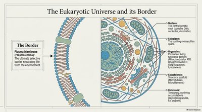
Plasma Membrane: Composed of a lipid bilayer with embedded proteins, carbohydrates, and cholesterol.
Functions: Selective transport, communication, anchoring, and identity.
Membrane Thickness and Microscopy
The plasma membrane is only 7.5 nm thick, making it invisible to light microscopy. Its true structure is revealed by electron microscopy, which shows a double-line appearance.
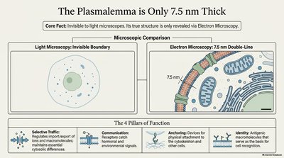
Selective Traffic: Regulates import/export of molecules.
Communication: Receives signals and hormones.
Anchoring: Provides attachment for cytoskeleton and extracellular matrix.
Identity: Antigens and markers for self-recognition.
The Fluid Mosaic Model
Membrane Structure and Dynamics
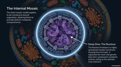
The fluid mosaic model describes membranes as dynamic, not static, with proteins and lipids moving within a flexible bilayer. Membrane proteins are distributed like icebergs in a sea of lipids, and the bilayer is asymmetrical.
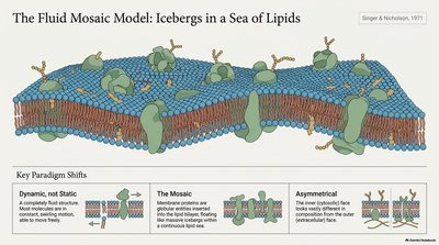
Dynamic: Lipids and proteins move laterally.
Mosaic: Proteins are interspersed among lipids.
Asymmetrical: Different faces of the bilayer have distinct compositions.
Anatomy of a Phospholipid
Phospholipids are the fundamental building blocks of membranes, featuring a hydrophilic head and hydrophobic tails. Their amphipathic nature drives membrane formation.
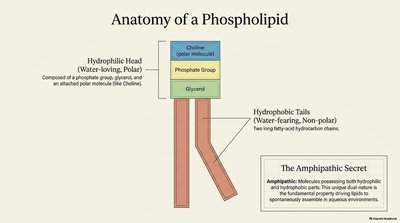
Hydrophilic Head: Polar, water-loving, contains phosphate and choline.
Hydrophobic Tails: Non-polar, water-fearing, composed of fatty acid chains.
Amphipathic: Possesses both hydrophilic and hydrophobic regions.
Spontaneous Self-Assembly
Phospholipids spontaneously assemble into bilayers in aqueous environments due to their amphipathic properties. This self-assembly is essential for cellular structure.
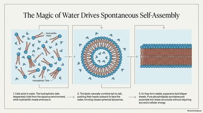
Hydrophobic Effect: Drives tails inward, heads outward.
Liposomes: Closed bilayer structures formed in water.
Membrane Fluidity and Lipid Movement
Membranes are dynamic 2D fluids, with lipids exhibiting lateral diffusion, flexion, and rotation. Flip-flop movement is rare and energetically unfavorable.
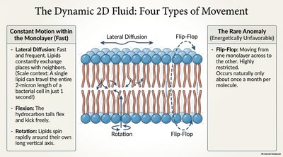
Lateral Diffusion: Lipids move rapidly within the monolayer.
Flexion: Hydrocarbon tails flex.
Rotation: Lipids spin around their axis.
Flip-Flop: Rare movement across the bilayer.
Saturated vs. Unsaturated Tails
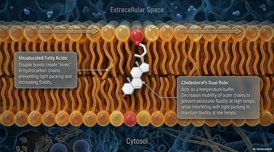
The fluidity of membranes is influenced by the saturation of fatty acid tails. Saturated tails are stiff and viscous, while unsaturated tails are fluid and loose.
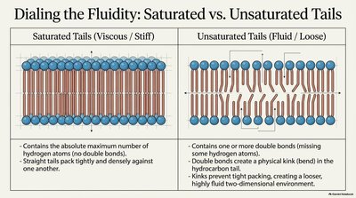
Saturated Tails: No double bonds, tightly packed.
Unsaturated Tails: Double bonds create kinks, increase fluidity.
Cholesterol: The Structural Mortar
Cholesterol is an amphipathic molecule that constitutes about 20% of animal cell membrane lipids. It acts as a structural mortar, filling gaps and modulating membrane fluidity.
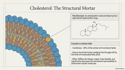
Function: Stiffens bilayer, reduces permeability, acts as a temperature buffer.
Lipid Asymmetry
Asymmetrical Distribution of Lipids
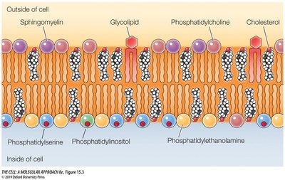
Membranes display asymmetry in lipid composition between the inner (cytosolic) and outer (noncytosolic) faces. Glycolipids are found exclusively on the outer face, while certain phospholipids are enriched on the inner face.
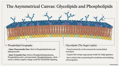
Phospholipid Geography: Outer face rich in phosphatidylcholine and sphingomyelin; inner face rich in phosphatidylethanolamine, phosphatidylserine, and phosphatidylinositol.
Glycolipids: Sugar-lipids found on the outer face, important for cell recognition.
Membrane Proteins
The Functional 50%
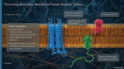
Proteins make up about 50% of the membrane mass and are responsible for most cellular functions. They are classified as integral or peripheral based on their association with the bilayer.
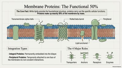
Integral Proteins: Permanently embedded, span the bilayer.
Peripheral Proteins: Temporarily attached to one face.
Major Roles: Transporters, anchors, receptors, enzymes.
Membrane-Cytoskeleton Interactions
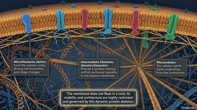
The membrane is supported by a dynamic protein skeleton, including microfilaments, intermediate filaments, and microtubules. These structures restrict mobility and provide architectural support.
Glycocalyx and Cell Surface Identity
The Glycocalyx: Cell's Sugar Armor
The glycocalyx is a carbohydrate-rich cell coat found on the exterior of the plasma membrane, formed by glycoproteins and glycolipids. It provides protection, adhesion, and can be a vulnerability point for pathogens.
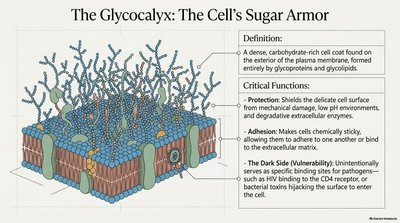
Protection: Shields cell surface from damage.
Adhesion: Enables cells to bind to each other or the extracellular matrix.
Vulnerability: Pathogens may exploit specific binding sites.
Blood Group Antigens and Membrane Identity
ABO Blood Group Antigens
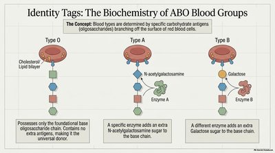
Blood group antigens are oligosaccharides linked to membrane lipids or proteins. The arrangement of sugar chains determines blood type and immune compatibility.
Antigenic Determinant: Carbohydrate portion of glycoproteins/glycolipids.
Immune Response: Transfused blood with different carbohydrate markers is recognized as foreign.
Structural Differences: O antigen is foundational; A antigen adds N-acetylgalactosamine; B antigen adds galactose.
Blood Type | Antigen Structure | Enzyme |
|---|---|---|
O | Fucose, galactose, N-acetylglucosamine, glucose | All people have O antigen enzyme |
A | O antigen + N-acetylgalactosamine | Enzyme A adds N-acetylgalactosamine |
B | O antigen + galactose | Enzyme B adds galactose |
Summary Table: Membrane Components and Functions
Component | Structure | Function |
|---|---|---|
Phospholipids | Bilayer, amphipathic | Barrier, fluidity |
Cholesterol | Rigid steroid rings | Stiffens, buffers fluidity |
Proteins | Integral/peripheral | Transport, signaling, anchoring |
Glycolipids | Sugar-lipids | Cell recognition |
Glycocalyx | Carbohydrate coat | Protection, adhesion |
Key Equations and Concepts
Amphipathic Nature of Phospholipids
The amphipathic property is essential for bilayer formation:
Membrane Fluidity
Fluidity is influenced by temperature, cholesterol, and fatty acid saturation:
Blood Group Antigen Structure
Antigenic determinants are defined by specific oligosaccharide additions:
Conclusion
Cellular membranes are complex, dynamic structures essential for compartmentalization, communication, and identity. Understanding their composition, fluidity, and surface markers is fundamental to cell biology and biomedical science.
